MAGNESIUM DEFICIENCY IN THE PATHOGENESIS OF DISEASE
Early Roots of Cardiovascular, Skeletal
and Renal Abnormalities
Goldwater Memorial Hospital
New York University Medical Center
New York, New York
1980
(include the word "jacket" to search only in this book)
| Jacket | Preface | Contents | Introduction (Chapter 1) |
Chapter: | 2 | 3 | 4 | 5 | 6 | 7 | 8 | 9 | 10 | 11 | 12 | 13 | 14 |
| Appendix | Bibliography (A-D), (E-K),
(L-R), (S-Z) |
Part I: Chapter 4
MAGNESIUM DEFICIENCY DURING GESTATION, INFANCY, AND EARLY CHILDHOOD
The magnesium levels at birth (indicated by cord levels) reflect the fetal response to maternal conditions during gestation: systemic and placental, and the ease or difficulty of delivery with resultant normal or hypoxic state of the newborn infant. Conditions that lead to neonatal hypermagnesemia might mask an underlying magnesium deficiency. Hypermagnesemia might result from administration of pharmacologic doses of magnesium to the mother shortly before delivery for management of toxemia of pregnancy, or from egress of magnesium from the tissues of infants subjected to anoxia, acidosis, or surgery. Exchange transfusions with citrated blood profoundly affect magnesium as well as calcium homeostasis. Levels during the first week of life reflect the infant's adjustment to independent life in the absence of immediate maternal blood-borne factors, and are affected by the nature of milk and supplements provided. The nature of feeding also influences levels later in infancy. Metabolic abnormalities that interfere with magnesium absorption or retention, although not common, have produced severe mineral imbalances that have focused pediatricians' attention on magnesium. More common conditions, such as severe diarrhea and intestinal malabsorption syndromes, which also cause hypomagnesemia, have further stimulated the pediatrician to be alert to magnesium loss. This section calls attention to the conditions and mechanisms that make infants susceptible to magnesium deficiency and presents speculations as to possible late, as well as overt, immediate sequellae.
4.1. Infantile Magnesium Deficiency: A Factor in Hypocalcemic Tetany, Seizures, and Respiratory Distress
It has long been recognized that neonatal hypocalcemia causes neuromuscular irritability and frank seizures. That the hypocalcemia is secondary to hypomagnesemia in many instances is now clearly established: as a factor in neonatal hypo parathyroidism, in vitamin-D-resistant rickets, and in genetic magnesium malabsorption. Treatment of infantile hypocalcemia with calcemic agents, which can intensify any preexisting magnesium insufficiency, has been shown to cause severe hypomagnesemia and intensification of the clinical manifestations that predicated their use. It is possible that such treatment can be a contributory factor in subsequent renal tubular wasting of magnesium, which can result from intraluminal renal tubular calculi.
Acute magnesium deficiency of infancy severe enough to cause tetany or convulsions, usually in association with hypocalcemia and occasionally with hypercalcemia, was first reported in 1921 by Denis and Talbot. They analyzed plasma calcium levels in 116 hospitalized infants and young children and reported magnesium levels in 38 of those patients. Of the 24 who had hypomagnesemia, six had seizures; two of the older children, four and five years of age, who had been diagnosed as having epilepsy or petit mal had hypocalcemia as well as hypomagnesemia. Three more had tetany; one of those died with laryngospasm at seven months. There were four additional young children (seven months to three years of age) with convulsions, and one with tetany, who had not had their plasma magnesium levels measured. One with microcephaly and mental retardation and one with mental retardation alone had plasma calcium levels of 9.2 and 9.7 mg/100 ml at seven months and two years, respectively. (Another baby with microcephaly and mental retardation, who had plasma calcium of 13.5 mg/100 ml at one year of age, may be the first recorded instance of the infantile hypercalcemia syndrome.) The remaining three babies with seizures or tetany had plasma calcium levels between 5.5 and 8.2 mg/100 ml.
Until the past 15 years, few papers evaluated the magnesium status of infants with abnormalities that later investigations suggest might well have been related to perinatal magnesium deficiency. The infants with tetanic or convulsive signs of hypocalcemia, which were associated with maternal hyperparathyroidism and became worse following treatment with calcemic agents, might have had contributory magnesium deficiency. So, also, might those born after complicated pregnancies or difficult deliveries, which has been shown to predispose to infantile convulsions (S. Wallace, 1972).
The role of hypomagnesemia in infantile convulsions has gained increasing recognition since J. A. Davis et al. (1965) reported an infant with hypomagnesemic neonatal fits, born to a mother with chronic malabsorption, and Paunier et al. (1965, 1968b) identified isolated magnesium malabsorption of infancy as a newly recognized genetic disorder. This condition is associated with hypocalcemic tetany and convulsions that require high doses of magnesium for correction. Use of calcium infusions or calcemic agents, such as high doses of vitamin D or parathyroid hormone, can intensify the neuromuscular irritability, and often do not even correct the hypocalcemia. However, far more infants than those unusual children with magnesium malabsorption are subject to hypomagnesemia. For example, the same year that Paunier et al. (1965) published their preliminary report, Davis et al. (1965) reported an infant boy with convulsions that started on the eighth day of life, and who had hypocalcemia, hypomagnesemia, and hypoglycemia. His intermittent fits became continuous following glucose and calcium infusions that raised his blood glucose to normal but exerted no influence on the hypocalcemia (Fig. 4-1). The seizures stopped within 30 seconds of intravenous administration of 2.5 mEq of magnesium, and his strongly positive Chvostek's sign became negative. The authors considered maternal hyperparathyroidism (secondary to long-term intestinal malabsorption) to have resulted in transitory suppression of her baby's parathyroid function. He responded to PTH by increased clearance of phosphate and decreased calcium and magnesium excretion, despite which his serum magnesium again declined, but without recurrence of convulsions.
Following the detailed study of the second reported (male) infant with magnesium malabsorption (Salet et al., 1966), and the suggestion that the disease might be hereditary in a third boy (M. Friedman et al. 1967), two more male infants developed convulsive hypomagnesemic hypocalcemia. One was born to a mother with poorly controlled diabetes mellitus (Clarke and Carré, 1967) and thus might have had intrauterine magnesium deficiency. The other was born to a mother with hypophosphatemia, who had received Dilantin therapy for many years (Dooling and Stern, 1967), and thus might have been magnesium deficient before and after birth. The infant born to the diabetic mother (Clarke and Carré, 1967) had had a low Apgar score at one minute and developed respiratory distress a few hours after birth. He had clonic convulsive movements on day 13, which responded to addition of calcium chloride to his formula until day 32, when his convulsions recurred. They intensified on addition of AT-10 (a dihydrotachysterol), high dosage vitamin D, and intravenous calcium gluconate, which did not increase his serum calcium levels. His serum magnesium was then measured and found to be 0.6 mEq/liter. A single intramuscular injection of magnesium (1 ml 50% MgSO4 resulted in cessation of convulsive movements a few minutes after the injection; the improvement persisted thereafter and no further magnesium supplements were given. The infant who had received the exchange transfusion (Dooling and Stern, 1967) showed continuation of irritability, tremulousness, and convulsions, after a focal seizure on day 6, that persisted (during calcium therapy) until his hypomagnesemia was detected and corrected. Atwell (1966) presented detailed studies of three infant boys who developed hypomagnesemia and hypocalcemia and were unresponsive to calcium infusions after neonatal gastrointestinal surgery, but who responded to magnesium (Fig. 4-2).
The clustering of reports of neonatal infants, whose hypocalcemic convulsions could be directly attributed to magnesium deficiencies of different etiologies led to an editorial (Canad MAJ, 97:868, 1967) that pointed out that hypomagnesemia is more likely to be a crucial medical problem than a chance occurrence. Stressed was the need for ready availability of facilities to monitor serum magnesium levels, certainly in convulsing infants, and also in other conditions associated with hypomagnesemia, including hypervitaminosis D and use of diuretics, and in the protein-calorie-malnutrition syndrome. Because of sudden death occurring in infants receiving exchange transfusions, and the evidence that citrated blood lowers blood magnesium levels (Bajpai et al., 1967a,b), the editor also called for determinations of magnesium levels in such infants, or preferably using heparinized rather than citrated blood for exchange transfusions. Neonatal infants requiring major surgery, who also generally are transfused, are also at risk of hypomagnesemia (Atwell, 1966; Jalbert et al., 1969).
There have been many published case reports and reviews published since, in which hypomagnesemia is the common denominator in several otherwise unrelated conditions characterized by neonatal and later infantile tremors, tetany, and convulsions. Most are associated with hypocalcemia, but several show a poor correlation with plasma calcium levels. Whether hypocalcemic tetany or convulsions associated with normal magnesium levels in the serum (which can rapidly attain normal levels despite tissue deficit) is another manifestation of a related metabolic disorder requires further study.
4.1.1. Magnesium Deficiency in Metabolic Convulsions of Otherwise Normal Newborn Infants
The group in Scotland that considers disturbed magnesium metabolism to play a significant role in neonatal convulsions in otherwise normal infants (Forfar et al., 1971/1973; J. K. Brown et al., 1972; Cockburn et al., 1973; Forfar, 1976; T. Turner et al., 1977) observes that this syndrome occurs in bottle-fed, but generally not in breast-fed, infants. They have presented evidence that both plasma and cerebrospinal fluid (CSF) levels of magnesium and calcium are lower in convulsing than in normal infants; the CSF phosphorus level of convulsing infants is normal despite hyperphosphatemia. The babies with convulsions are described as classically "jittery." They found the syndrome to be severe in 35% and lesser in degree more frequently. Among 75 consecutive newborn infants with convulsions considered due primarily to disordered mineral metabolism, seen over a two-year period, subnormal calcium levels (more than 2 S.D. below the mean) were seen in 92%, subnormal magnesium levels in 52%, high phosphorus levels in 67%, and combinations of biochemical disturbances in 80% (Fig. 4-3). Hypocalcemia was associated with hyperphosphatemia in about 60% and with hypomagnesemia in about half of the cases. Hypomagnesemia without hypocalcemia was seen in 7%, almost half of whom also had normal phosphorus levels. Convulsions considered due primarily to brain damage (in 60 additional infants) often also exhibited mineral metabolism derangement, predominantly hypocalcemia and hyperphosphatemia (J. K. Brown et al., 1972). Infants fed evaporated milk formulas had low magnesium and high phosphorus levels, comparable with levels of convulsing infants in 68 and 80% of the controls. In an evaluation of clinical and chemical relationships in neonatal convulsions, the group (J. K. Brown et al., 1972) commented that they had encountered convulsions in 1.4% of live-born infants. Most of those classified as due to brain damage occurred in the first three days of life; most of those considered metabolic in origin occurred from the fourth day on (Fig. 4-4). They noted that the proportion of metabolic to brain-damage convulsions seems to have risen markedly in reports published since 1969, as compared with reports published between 1954 and 1960, during which time brain-damage-induced convulsions were predominant. Since metabolic convulsions are more amenable to correction, this is an important point in terms of management of convulsing infants.
Wong and Teh (1968) had earlier reported hypomagnesemia without hypocalcemia in five otherwise normal infants, during the week after birth. (This was part of a study of 40 babies and young children with convulsions, tremors, or muscular twitchings, 13 of whom had hypomagnesemia alone, and 27 of whom also had hypocalcemia). When symptoms were present, both total and ultrafiltrable mean levels of magnesium were significantly lower than in controls (p = < 0.001). The major decrease was in the ultrafiltrable moiety. Keen (1969), like Forfar and his colleagues (supra vide) called attention to the increasing incidence of infantile convulsions of metabolic origin in England. Of 100 infants with seizures in the first 4 weeks of life, 36 had hypocalcemia, with peaks of incidence in the first 48 hours and between the 4th and 10th days of life. Only toward the end of this 23-month study were magnesium levels determined. Details of the inconstant association of hypomagnesemia with hypocalcemia were not given, but the investigator considered the response of refractory hypocalcemic fits to magnesium (Davis et al., 1965) as suggestive of its importance in this syndrome. He, too, commented on the disproportionate distribution of convulsions among bottle-fed as compared with breast-fed infants. Harvey et al. (1970) also showed that the mean magnesium level was lower in bottle-fed than breast-fed infants by the seventh day of life, and that among those with convulsions the mean was even lower. In this series, even many of the nonconvulsing infants had hypomagnesemia and hypocalcemia. This recalls Bruck and Weintraub's (1955) admonition that asymptomatic hypocalcemia should not be considered "physiologic," since transition from latent to manifest tetany is frequent and can occur unexpectedly. The same is likely to be true for hypomagnesemia. Furthermore, because of the evidence that prolonged chronic magnesium deficiency can contribute to cardiovascular, renal, and bone abnormalities, overt symptomatology may not be the major risk.
The infant reported by Vainest et al. (1970) might be an example of delayed as well as acute complications of magnesium deficiency of infancy. Although this infant had not had his severe hypomagnesemia (0.4-0.7 mEq/liter) detected until three days before he died at five and a half months, there is strong inferential evidence that magnesium deficiency was likely to have played a contributory role. He was the ninth child of a woman who had been treated for tuberculosis, and thus was probably magnesium depleted. [High parity contributes to the magnesium drain on the mother, and aminoglycoside antibiotics are magnesium wasters (Vanasin et al., 1972).] Five of her seven sons had had seizures; three died. Two, counting the propositus, whose hypomagnesemia had been identified late (after massive calcemic therapy), had arterial calcinosis. That infant also had renal and myocardial calcinosis.
The frequency of low magnesium levels among infants with symptomatic hypocalcemia was noted by the investigators cited above, and in subsequent studies. Stern and Harpur (1971/1973) briefly reported six newborn infants whose hypocalcemia was clearly secondary to their hypomagnesemia. Radde et al. (1972) commented that symptoms and signs attributable to low ionized calcium levels were found only in infants who had low plasma levels also of magnesium. Tsang (1972), who reviewed in detail the factors contributing to neonatal magnesium disturbances, also commented on the concomitant hypocalcemia, and vice versa. Subsequent work from his group has elucidated the infants at greatest risk of the combined divalent cation deficiencies (Tsang et al., 1973, 1974, 1976, 1977a,b; Tsang and Brown, 1975, 1977). David and Anast (1974) found that plasma magnesium levels were significantly lower in hypocalcemic than in normal or sick neonates.
Most of the infants described in this section were newborn. Convulsions and tetany associated with hypomagnesemia have also been reported in older infants and young children. Febrile convulsions are frequently associated with lower than normal serum magnesium levels, often without hypocalcemia (Chhaparwal et al., 1971). A "meningo-encephalitic, or tremor" syndrome in Indian children has also been associated with hypomagnesemia in infants of 6-24 months of age, who have evidence of mental retardation and malnutrition (Chhaparwal et al., 1971/1973). Severe magnesium deficiency also occurs during repair of protein calorie malnutrition (see pp. 122-128).
4.1.2. Low-Birth-Weight Infants
Lower cord blood magnesium levels have been reported in low-birth-weight infants than in full-term infants (Breton et al., 1960; Review: Ferlazzo and Lombardo, 1971). When the low birth weight is due to prematurity, the low cord blood levels can be attributed to the subnormal accumulation of minerals in the final weeks of gestation. Widdowson and Dickerson (1962), who have tabulated the mineral contents of 1.5 kg, 2.5 kg, and full-term babies, have shown that the magnesium content of the more immature or smaller babies is only 42% that of normal size infants, while that of 2.5-kg infants is 76% that of the normal full-term baby. In regard to the tendency toward hypocalcemia of premature infants, the 1.5- and 2.5- kg infants have 36% and 68% the calcium contents, respectively, of full-term babies. This study did not differentiate between immature infants and those that are small for gestational age (SGA) as a result of intrauterine growth retardation (IUGR).
The hypocalcemia and hypomagnesemia of low-birth-weight infants can reflect inadequate stores accumulated before birth, in addition to postnatal problems in homeostasis. Their hyperphosphatemia can derive from tissue breakdown and be aggravated by inappropriate dietary intakes, functional immaturity, and hormonal imbalances. The hyperphosphatemia associated with hypocalcemia and hypomagnesemia that is found in full-term infants fed cows' milk rather than breast milk and that is aggravated by vitamin D is further discussed on pp. 105-108.
Renal tubular immaturity has been proposed as an explanation of the inability of the neonate to eliminate excess phosphate, whether endogenous or exogenous, that is associated with persistent hypocalcemia and hypomagnesemia. Rubin et al. (1949) showed that aspects of renal function mature at different rates, usually reaching adult values during the second year of life. Dean and McCance (1948) and L. Gardner et al. (1950) reported that renal tubular immaturity was responsible for the low phosphate clearance that they reported in neonatal infants. This fits the experimental evidence suggesting absence of fetal phosphaturic response to exogenous PTH (Garel and Barlet, 1974). Tsang et al. (1973b) found that phosphorus excretion increased in premature infants over their first three days of life, whether or not PTH was given. Their fractional tubular reabsorption fell and there was no significant difference in phosphorus excretion or reabsorption between the PTH-treated and nontreated infants.
The theory that transient hypoparathyroidism of infancy is a result of parathyroid immaturity has been discussed earlier. If valid, this theory is even more applicable to low-birth-weight infants and might explain their subnormal PTH response to neonatal hypocalcemia. Also suggested frequently is the possibility that fetal hypercalcemia, possibly deriving from maternal hyperparathyroidism-induced hypercalcemia, might cause fetal PTH suppression, mediated by resultant fetal hypercalcemia (Review: Tsang et al., 1976b). However, cited experimental studies have shown that experimental dietary- or hyperparathyroidism-induced calcium and phosphate aberrations are not reflected by parallel changes in the fetal blood. Fetal parathyroids function to maintain the calcium homeostasis. Furthermore, hypercalcemia during late gestation is uncommon even in the presence of "physiologic" hyperparathyroidism. Thus, it seems plausible that it is not parathyroid immaturity but postnatal factors that prevent normal PTH reactivity. For example, hypocalcemic hyperphosphatemic premature infants have responded to injections of exogenous PTH with transient rises in serum calcium and magnesium in the first few days of life (Tsang et al., 1973; David and Anast, 1974; Root et al., 1974), indicating that there was bone mineral mobilization in response to PTH (Fig. 4-5, Tsang et al., 1973a), even in infants born prematurely.
In the case of infants with IUGR, such as are commonly born to mothers with toxemias of pregnancy and to young primiparous mothers, significantly lower levels of serum magnesium have been detected than in other low-birth-weight infants (Fig. 4-6, Tsang and Oh, 1970: Jukarainen, 1971). Tsang and Oh (1970) suggested that the low serum magnesium levels in IUGR infants might reflect disturbed placental transfer of magnesium or abnormal fetal magnesium metabolism as part of the intrauterine malnutrition syndrome. Hypocalcemia has been shown to be more striking than hypomagnesemia in IUGR neonates (Tsang et al., 1975). Such neonatal hypocalcemia in infants with placental insufficiency has been associated with impaired transfer of calcium from mother to fetus (Khattab and Forfar, 1971). Tsang et al. (1975) suggest that their findings (Tsang et al., 1973a,b, 1974) point toward shortened gestational age or birth asphyxia as more likely explanations of the disturbances in calcium homeostasis during the early neonatal period. The greater tendency of IUGR infants than full-term infants to have poor bone mineralization and spontaneous bone fractures suggests that maintenance of divalent cation homeostasis in utero might be achieved by hyperactivity of fetal parathyroids in response to intrauterine malnutrition, when there is faulty placental transport of calcium and magnesium from maternal to fetal circulation.
The observation that IUGR infants often exhibit neonatal hyperirritability and jitteriness (Michaelis et al., 1970; Ferlazzo and Lombardo, 1971; Tsang et al., 1975) suggests that, in addition to hypocalcemia, magnesium deficiency also be considered. The failure to find hypomagnesemia at 4 hours, and its decline by 24-48 hours, especially in infants whose hypocalcemia also becomes more notable at that time (Fig. 4-7, Tsang et al., 1975b), suggests that hypoxia at birth, which is common in IUGR infants (Tsang et al., 1975), can be contributory and might mask the magnesium deficiency. Serum magnesium values being a poor index of tissue magnesium status, percentage retention of magnesium-load tests might prove a more valid means of ascertaining whether the irritability of IUGR infants can be partially attributed to magnesium deficiency (Harris and Wilkinson, 1971; Caddell, 1975).
4.1.3. Neonatal Hypoxia
Infants born after difficult deliveries and who have birth apnea have been found to have hypermagnesemia shortly after birth (Engel and Elm, 1970). It is probable that the source of this elevated serum magnesium is from the tissues, injured as a result of the hypoxia, as has been demonstrated in war injuries and clinical or experimental shock (Beecher et al., 1974; Root et al., 1947; Canepa and Gomez-Pavira, 1965; W. Walker et al., 1968; N . Goldsmith et al.,1969; Flynn et al., 1973, 1976/1980). The accompanying acidosis enhances the shift of bone minerals to the extracellular space (Barzel and Jowsey, 1969; Raisz, 1970). Thus, such infants, despite their transient hypermagnesemia or normal magnesium levels (Fig. 4-8) (Tsang et al., 1974), may actually suffer from body depletion of magnesium. Their drop in serum calcium in the first few days of birth has been generally blamed for the hyperirritability, jitteriness, convulsions, and periods of apnea, common in hypoxic infants (Oppé, 1970). However, they frequently also show as striking depressions in their serum magnesium levels and a lesser drop in serum phosphorus (Fig. 4-9, Tsang et al., 1974). The rise in serum phosphorus, which precedes the rises in the divalent cations, suggests that PTH-mediated mobilization of bone mineral might not then be operative. The rise in serum phosphorus can be caused by several factors. The initially higher than maternal values might be endogenous in that it is caused by endogenous tissue breakdown, which is associated with stress of delivery and birth asphyxia. The subsequent rise might derive from bone mineral efflux, high phosphate intake (from cows' milk), and renal tubular inability to eliminate the phosphorus load in the early days of life. Asphyxiated infants, whose serum magnesium levels dipped only slightly at 12 hours and then rose to normal by 24 hours, were compared with asphyxiated infants whose hypoxemia (starting at 12 hours) persisted through 48-72 hours (Tsang et al., 1974). The hypocalcemia of the latter group was more profound, and correction of acidosis took longer than it did in the asphyxiated infants with normal serum magnesium levels. The drop in serum magnesium levels within 12-24 hours after asphyxia may well reflect the low reserves of magnesium in neonatal infants, or the shift from extracellular to intracellular space on correction of the hypoxia and acidosis.
4.1.4. Neonatal Infants of Diabetic Mothers
Infants of diabetic mothers can either be premature or large for gestational age, often exhibit respiratory distress and acidosis, and also frequently show rising serum phosphorus and falling serum calcium and magnesium levels by 24-48 hours after birth (Fig. 4-10, Tsang et al., 1972). This had been speculated to reflect maternal hyperparathyroidism of diabetic mothers. However, Tsang et al. (1972) noted that diabetic mothers had serum calcium levels within normal limits. Since they did not have hypercalcemia, suppression of fetal parathyroids from this source seems questionable. Functional hypoparathyroidism of the infants was considered unlikely when they were found to exhibit short-term calcemic response to PTH injections (Fig. 4-11), indicating bone mineral mobilization. Although administration of PTH to infants of diabetic mothers caused more phosphaturia than was seen in nontreated infants of diabetic mothers, there was no difference in percentage tubular reabsorption of phosphorus in the two groups, suggesting renal immaturity. Their subsequent work showed no significant difference in serum PTH or total or ionized calcium levels in diabetic than in normal mothers (Tsang et al., 1975). Since they found that PTH levels of cord blood of infants of diabetic mothers (IDM) were not significantly lower than were those of controls, they assumed that the parathyroids of the IDM functioned as did those of normal infants. The observation that there was no significant increase in PTH levels in response to significant decreases in total and ionized calcium led Tsang et al. to assume a failure of production of PTH. Prematurity (9 of 13 infants of insulin-dependent mothers with gestational ages of 37 weeks or less), birth asphyxia (10 of the 28 IDM had 1 minute Apgar scores of 6 or less), and increased calcitonin secretion were also considered as possible explanations for the sustained hypocalcemia of the infants of diabetic mothers. The changes in IDM serum magnesium were not considered significantly different from those of controls in that study. However, although the maternal serum magnesium levels were within the same range in control and diabetic mothers, it is of interest that the cord blood levels of the normal infants, which were low, rose to about 1.7 mEq/liter by 76-96 hours, whereas the mean values of infants of insulin-dependent mothers remained about 1.5 mEq/liter. Their range of values at 24-48 hours was 1.35-1.7 mEq/liter and at 72-96 hours was about 1.4-1.5 mEq/ liter. The following year, Tsang et al. (1976b) reported that 21 of 56 IDM had serum magnesium levels at or below 1.25 mEq/liter on at least one occasion during the first three days, and that they did not exhibit the normal increase with postnatal age seen in normal infants. Subnormal neonatal serum magnesium levels were related to the degree of severity of diabetes, youth of the mothers, lower gravidity, and prematurity. Lower concentrations of serum magnesium were associated with less increase (or actual decreases) in serum concentrations of PTH from 48-72 hours, and conversely serum concentrations of magnesium at 72 hours were related to parathyroid function from birth to 24 to 48 hours of age (Fig. 4-12, Tsang et al., 1976c). Since diabetes mellitus is recognized to cause magnesium deficiency without the added requirements caused by pregnancy, it is not surprising that infants of diabetic mothers are particularly subject to magnesium deficiency. The interrelationship of their magnesium inadequacy, phosphate excess, and hypocalcemia with their parathyroid malfunction is an important clue to the complex hormonal/mineral interrelationships that may be mediated by a fundamental magnesium deficit.
4.1.5. Neonatal Hypermagnesemia
Hypoxia has been shown to cause loss of magnesium from tissues with resultant elevation of serum magnesium levels. Studies of serum from venously occluded arms (Whang and Wagner, 1966; S. P. Nielsen, 1969) have shown that even short periods of hypoxia cause egress of magnesium from the cells to the blood. Thus, it is not surprising that infants born after difficult deliveries and with birth asphyxia have had elevated serum magnesium levels at birth and shortly thereafter (Engel and Elm, 1970; Donovan et al., 1977b). Such infants, however, often exhibit hypomagnesemia within 12 hours after birth (Tsang et al., 1974), possibly reflecting inadequacy of tissue stores or the shift of extracellular magnesium to the intracellular phase with normal oxygenation.
Acidosis, common in low-birth-weight infants, is another cause of neonatal hypermagnesemia. Even minor drops of muscle pH (to 6.8) has been shown in vitro to cause significantly decreased muscle magnesium content (Gilbert, 1961). A clinical reflection of this observation is the hypermagnesemia of decompensated diabetic acidosis (Marlin et al., 1958). Thus, the normal or elevated serum magnesium seen in acidotic infants immediately after birth, despite the evidence that such infants are at risk of hypomagnesemia, should come as no surprise. Even normal infants have acidosis, due to elevated maternal lactic acid levels and to the period of anoxia during birth (Acharya and Payne, 1965). The levels fall as oxygenation is established, normally reaching adult values after two days. Infants with respiratory distress have prolonged acidosis and anoxia, which militate against restoring tissue levels of magnesium. This set of circumstances is likely to mask the underlying magnesium deficiency when serum magnesium levels are relied upon to reflect the magnesium status.
The most intensive study found on the magnesium levels of the neonate (Jukarainen, 1974) demonstrates that high-risk infants with hypocalcemia (whom one would expect to have hypomagnesemia) are likely to have normal magnesium levels in the early hours to days after birth. This investigator correlated many factors that influence neonatal homeostasis, considering gestational and perinatal abnormalities. As many as nine blood samples were analyzed for Mg/Ca/P in the infants during the first five days of life. He found that these longitudinal studies showed that there was an inverse correlation between the serum magnesium and gestational age. The premature and low-birth-weight infants (who have been shown to be more susceptible to hypocalcemic tetany and convulsions) had essentially normal serum magnesium with their hypocalcemia in the first five days, as compared with full-term infants whose hypocalcemia correlated positively with hypomagnesemia during the same period. Infants of diabetic mothers also showed relatively higher serum magnesium levels, in association with their hypocalcemia during the first few days, but the magnesium levels tended to drop toward the end of the observation period. Jukarainen (1974) concluded that the inverse relationships between calcium and magnesium levels in the early days of life of the high-risk infants probably reflected disturbed magnesium homeostasis (such as has been seen with hypoxic and acidotic egress of magnesium from the cells).
Direct evidence that this might explain the above findings was provided by Yamashita and Metcoff (1960), who found that the skeletal muscles of premature infants were edematous, and that the levels of normal intracellular cations and of magnesium-dependent enzymes were significantly lower than normal. Chiswick (1971) also noted edema in hypocalcemic neonatal infants, and noted that the serum magnesium levels of the hypocalcemic infants were higher in infants with edema than in those without.
Infants born to mothers given pharmacologic doses of magnesium for eclampsia shortly before delivery have been born with hypermagnesemia and secondary respiratory depression, areflexia, and paralysis (Fishman, 1965; Brady and Williams, 1967; Lipsitz and English, 1967; Lipsitz, 1971). Serum levels as high as 15 mEq/liter were detected in one such infant, who recovered following treatment by exchange transfusion (Brady and Williams, 1967). However, Lipsitz (1971) found no correlation between (1) the cord or newborn serum magnesium levels and the Apgar score; (2) the total dose of magnesium given to the mother and her serum magnesium level at delivery, or that of the cord blood; and (3) the total dose of magnesium and clinical evidence of neonatal magnesium toxicity.
Unlike adults, who excrete infused magnesium rapidly (Chesley and Tepper, 1958), neonates have a very low magnesium excretion rate (Lipsitz, 1971; Tsang, 1972). During the first few days of life, glomerular filtration rates are low (less than 0.34 mg/kg/24 hours); in premature infants the glomerular filtration rate and magnesium excretion is even less than in full-term infants (Tsang, 1972). Thus, it is not surprising that it has taken up to five days for neonatal hypermagnesemia to fall to normal levels (Lipsitz, 1971). Despite sustained elevated serum magnesium levels in infants born to toxemic mothers, given large amounts of magnesium for different periods of time before delivery, there have been surprisingly few instances of serious manifestations of hypermagnesemia. For example, only 8 of the 118 infants born to mothers given 30-40 g of magnesium sulfate i.m. during the 24 hours before delivery, had Apgar scores of 5 or less; none had cord magnesium levels above 6 mEq/liter during labor; no detectable adverse effects attributable to the magnesium alone were detected (Hutchinson et al., 1963).
The meconium plug syndrome, attributed to hypermagnesemic suppression of peristalsis, has been reported in two infants born prematurely to two eclamptic young women given high-dosage magnesium therapy shortly before delivery (Sokal et al., 1972). The cord blood serum magnesium level was 8.3 mEq/liter in the infant from whom it had been obtained; it was 6.0 mEq/liter at 3 hours of age in the other. It had dropped to 5.4 mEq/liter by 6 hours, 4.3 at 55 hours, to 4.3 mEq/liter in the first infant, and to 4.2 mEq/liter at 10 hours in the second. Neither had hypocalcemia at any time tested. Since epsom salt enemas have been known since the turn of the century to cause magnesium toxicity in children and adults (C. Fraser, 1909; Fawcett and Gens, 1943), this treatment of hyaline membrane disease, which has led to fatal consequences of severe hypermagnesemia, is no longer recommended (Tsang, 1972; Outerbridge et al., 1973).
4.1.6. Magnesium Depletion by Exchange Transfusions with Citrated Blood
Exchange transfusions with blood to which acid-citrate-dextrose (ACD) solution has been added are known to cause infantile hypomagnesemia (Dooling and Stern, 1967; Bajpai et al., 1967a,b; Z. Friedman et al., 1971,1972). Although it has long been known that weakly dissociated salts of citrate are formed with both magnesium and calcium (Hastings et al., 1934; Walser, 1961), and citrate infusions to dogs have caused both hypomagnesemia and hypocalcemia [total (Bunker et al., 1962) and ionized (Killen et al. 1971)], the customary procedure for infants receiving exchange transfusions who develop irritability, seizures, or cardiac arrhythmias (Dooling and Stern, 1967; Rosefsky, 1972) has been to provide calcium with the transfusion and to monitor the serum calcium levels. Generally, only when the condition fails to improve has the magnesium status been explored and magnesium therapy instituted. An editorial (Canad MAJ, 97:868, 1967) considered sudden death during the course of the exchange a possible consequence of the citrate-induced reduction in serum ionic magnesium. Two groups of investigators in Canada demonstrated that the serum ionic magnesium dropped substantially during exchange transfusion with ACD blood (Bajpai et al., 1967a,b; Z. Freidman et al., 1971, 1972). The first group (Bajpai et al., 1967b) noted electrocardiographic changes (flattening of T waves) when the serum Mg fell below 0.8 mEq/liter. The second group (Z. Friedman et al., 1972) considered it likely that the magnesium-responsive arrhythmia that developed during the fourth ACD plus calcium transfusion (Rosefsky, 1972) was likely to have reflected also a reduction in ionic calcium, despite the administration of calcium gluconate. A more detailed report (Radde et al., 1972), presented evidence that abnormal symptoms and signs are found almost exclusively in infants whose plasma levels of both cations were below the lower limits of normal. Recently Donovan et al. (1977a) showed that exchange transfusion (which they confirmed lowered serum levels of ionized magnesium and calcium) increases PTII levels, as measured by immunoassay.
An overload of citrate similar to that of the exchange transfusion of infants is the use of ACD blood prime in cardiopulmonary bypass procedures. Killen et al. (1972) showed that severe depression of ionized magnesium (Fig. 4-13A) could be prevented by adding magnesium sulfate: 3 ml of 10% solution per unit of ACD blood (Fig. 4-13B). Since low magnesium levels are common in patients to undergo open- heart surgery, magnesium therapy of such patients is often necessary (Holden et al., 1972; Khan et al., 1973).
When transfusions, using citrated blood, are given to those whose underlying condition makes, it likely that they might be magnesium deficient before the transfusion, severe depletion may ensue. Jalbert et al. (1969) reported such an instance in the case of a premature infant born to a preeclamptic mother. The infant developed mucoviscidosis and intestinal obstruction requiring resection, during which citrated blood transfusions were given. Calcium-refractory seizures developed that responded only to magnesium repletion.
4.1.7. Low Ionized Calcium and Hypomagnesemia
In view of the drop in ionized calcium and magnesium caused by citrated blood transfusions, attention should be paid to other more common conditions in which neonatal tetany has been correlated with decreased ionized calcium levels. (Ionized magnesium is less readily measured, and thus is rarely reported.) The possibility that asymptomatic neonatal hypocalcemia might be related to normal levels of ionized calcium despite low total calcium has long been suspected (Bruck and Weintraub, 1955) and more recently verified. Bergman (1972) showed that symptomatic neonatal hypocalcemia is associated with lower levels of ultrafiltrable fractions of calcium than of total calcium. On the other hand, D. M. Brown et al. (1972) measured the ionized fraction of calcium, and found no correlation between low ionized calcium levels and symptomatic hypocalcemia. Sorell and Rosen (1975) found symptoms with decreases in ionized calcium to a critical level of 2.5 mg 100 ml. Bergman (1974) showed that up to 10-12 hours after birth, the decrease in total calcium is mostly caused by a decrease in the ultrafiltrable fractions. Since symptomatic hypocalcemia seems to be better related to les decreases in ultrafiltrable calcium that consists of ionized calcium plus complexed calcium (about 14% of total calcium: Walser, 1961) than to the ionized fractions, it may be speculated that the change from asymptomatic to overt hypocalcemia might be contributed to by a drop in the complexed fraction. It can be presumed that the HPO4 fraction is unlikely to be low in a condition associated with hyperphosphatemia. The citrate fraction, which is dependent on vitamin D, seems a likely candidate for consideration. It is a complex question, however, since vitamin D deficiency (in rats) has been correlated with decreased blood and bone citrate levels (Harrison et al. ., 1957). Vitamin D administration to rachitic rats has raised the citrate levels (Steenbock and Bellin, 1953), but excess vitamin D (as in acute infantile hypercalcemia related to hyper reactivity to vitamin D) is associated with subnormal blood citrate levels (Forfar et al., 1959; Lindquist, 1962). Radde et al. (1972) found that, at least in newborn infants, symptomatic hypocalcemia only occurred when low ionized calcium levels were present with concomitant hypomagnesemia, an interesting observation in view of the vitamin D resistance of magnesium-deficient patients. Sorell and Rosen (1975), finding both normal and low serum magnesium levels in symptomatic hypocalcemia, did not confirm the report of Radde et al. (1972). However, of the seven infants and young children they reported, all but one (who had sepsis and thus might have had acidosis) had hypomagnesemia. The other two with normal serum magnesium levels in their series of nine were 17- and 19-year-old patients with renal failure, a condition that has been associated with tissue magnesium depletion despite even hypomagnesemia (Lim and Jacob, 1972c). One of the infants developed hypomagnesemia and hypocalcemia after cardiac surgery.
4.2. Treatment of Infantile Conditions Associated with Abnormalities of Magnesium
4.2.1. Correction of Neonatal Acidosis
When acidosis develops in the newborn infant, it is customary to treat it with sodium bicarbonate or sodium lactate. Unfortunately, the conditions that give rise to acidosis not infrequently are associated with magnesium egress from the cells. Infusions of sodium lactate cause substantially increased urinary output of magnesium (Barker et al. 1959). Thus, the production of negative magnesium balance in infants whose postoperative acidosis was thus corrected, and the production of hypomagnesemia (Atwell, 1966), is not surprising. (The stress of surgery also increases magnesium loss.) Correction of renal acidosis with lactate, citrate, or bicarbonate has also caused hypomagnesemia (Randall, 1969). Administration of sodium bicarbonate to acidotic neonatal infants has reduced serum ionic calcium levels (Radde et al., 1972; Tsang et al., 1977a,b) and has also lowered serum magnesium levels (Radde et al., 1972; Jukarainen, 1974). The higher the serum bicarbonate levels, the lower the serum magnesium levels (Jukarainen, 1974).
4.2.2. Intensification of Magnesium Deficiency by Treatment of Hypocalcemia with Calcemic Agents
It has been reiterated that infants with hypomagnesemia should not be treated with calcium or vitamin D (Tsang et al., 1977a; Seelig, 1978/1980). Nonetheless, since hypocalcemia is usually detected first in convulsing infants (magnesium determinations often being obtained only on failure of calcemic therapy to correct either the symptomatic or biochemical abnormalities), calcium alone or with vitamin D is still usually the first approach. In fact, prophylactic administration of calcium has been recommended for low-birth-weight or asphyxiated infants who are at particular risk of hypocalcemia (D. R. Brown et al., 1976; Salle et al., 1977). It is realized and cautioned that when symptomatic infantile hypocalcemia is found, hypomagnesemia should be sought (Editorial, Bruit. Med. J., 1973; Tsang et al., l977a). The observation that symptomatic infantile hypocalcemia develops almost exclusively when there is concomitant hypomagnesemia (Radde et al., 1972) lends support to the importance of seeking out a magnesium deficit. Since magnesium is predominantly an intracellular cation, and since levels in the blood are generally kept within narrow limits, relying on serum magnesium as the index of magnesium status of the body can give misleading information. This is particularly true for neonatal infants, whose serum magnesium can be elevated as a result of acidosis or asphyxia-induced egress of magnesium from tissue. The parenteral magnesium-load test is more reliable as a clue to magnesium depletion. For example, Harris and Wilkinson (1971) found that of nine infants suspected of magnesium deficiency, who had serum magnesium levels that were normal, four were deficient by the loading test, Byrne and Caddell (1975) found that there were infants in their survey whose magnesium deficiency would not have been detected by serum levels alone.
With high-risk infants, whose body stores of magnesium might be precariously low, it is possible that treatment directed toward correction only of hypocalcemia might thereby not only fail to correct convulsions, but might intensify occult cardiovascular and renal lesions. Such damage is caused by experimental magnesium deficiency, and is worsened by calcium, phosphate, and vitamin D excesses. Among infants with severe imbalances (low Mg/high Ca, P vitamin D intakes), the damage might be severe enough to cause acute and chronic signs and symptoms during infancy, leading to early death or chronic disorders that might be termed "congenital." Among those with less marked imbalances (i.e., whose prenatal stores were higher or whose postnatal calcemic challenges were less, there might be lesser degrees of damage that might lay the groundwork for adult cardiovascular and renal disease.
It seems likely, even though magnesium determinations had not been made, that the two infants described by D. Andersen and Schlesinger (1942) might have reflected the first of the two possibilities: convulsive hypomagnesemic hypocalcemia treated with calcemic agents, resulting in death in the fourth month of life. In addition to administration of calcium gluconate and moderately to extremely high doses of vitamin D (that lowered, rather than raised, the serum calcium levels) both infants were also given repeated blood transfusions for refractory anemia, and both were treated repeatedly for refractory acidosis. It is conceivable that the anemia was a sign of magnesium deficiency (Elm, 1973, 1976/1980). It is plausible that the calcium- and vitamin-D-refractory hypocalcemic neuromuscular irritability and seizures of both infants might have been caused by early magnesium deficiency that interfered with response to the calcemic agents, and that was intensified by that treatment and by the use of citrated blood for the anemia, and lactate and bicarbonate for the acidosis. One vomited several times daily and developed hypercholesterolemia; the other developed hypertension-all signs of vitamin D excess and in the case of increased blood pressure of a high Ca/Mg ratio. Both had peripheral and coronary arteriosclerosis; one had myocardial infarctions and the other had cardiomegaly. Both had severe renal damage: one predominantly fibrous replacement; the other (who had been given 300,000 IU vitamin D) also had renal calcinosis. Although their biochemical findings suggested hypoparathyroidism, they both had hypertrophied parathyroid glands and bone pathology, and were diagnosed at autopsy as having renal hyperparathyroidism. In view of the data reviewed in the foregoing section, the possibility that these infants had hyperparathyroidism secondary to magnesium deficiency, and that the deficiency interfered with the response of target organs to PTH (pseudohypoparathyroidism) or to vitamin D, and led to cardiovascular and renal disease should be seriously considered. Almost a quarter of a century later, severe hypomagnesemia (0.8 mEq/liter) was correlated with high-dosage vitamin D and calcium treatment of an infant whose hypocalcemic convulsions had started at one month (Salet et al., 1966), as in the prior two cases. Treatment with both cations was then instituted, with resultant elevation of low calcium levels to normal. Both hypocalcemia and hypomagnesemia (0.3 mEq/liter) recurred at three months, after treatment had been stopped. The baby again responded to combined cation therapy. When treatment was again stopped, he exhibited hyperphosphatemia, as well as hypocalcemia. PTH administration corrected the blood calcium and phosphorus levels, but lowered the blood magnesium level (0.5 mEq/liter). Vitamin D therapy again intensified the biochemical abnormalities and the convulsions. Like the infants described by Andersen and Schlesinger (1942) this infant's findings suggested hypoparathyroidism. However, his hypomagnesemia was identified early and treated intermittently until it became manifest that his vitamin-D-resistant hypocalcemia was secondary to magnesium malabsorption. This group found that high-dosage vitamin D increased his magnesium requirements and that treating with both magnesium and calcium was not as effective in raising cellular magnesium to normal levels as was treating with magnesium alone. They later found that this infant's magnesium malabsorption was familial, when a sibling was born with the same defect (Salet et al., 1970). High dosage vitamin D (100,000 IU daily) for familial hypoparathyroidism and convulsive hypocalcemia resulted in hypomagnesemia in a baby from a family with a high incidence of convulsions (Niklasson, 1970). This infant developed emotional lability and mental retardation, similar to that seen with hypervitaminosis D (Review: Seelig, 1969b). Her young sister later also developed hypomagnesemia. It was noted that infantile convulsions, with death during infancy (including one sudden unexplained death at four weeks), were common in the family of these sisters, whose parents were first cousins. The possibility that there was primary magnesium malabsorption or renal magnesium wasting in this family was not explored. The infant son (ninth child of a mentally retarded mother), who developed convulsions after three months of vitamin-D-supplemented (400 IU/day) dried milk formula, was the fifth son to develop seizures (Vainsel et al., 1970). Intravenous calcium gluconate and high dosage vitamin D (750,000 units per week) raised the serum calcium to low normal levels, but failed to control the seizures. Hypomagnesemia (0.4-0.7 mEq/ liter) was then identified, and magnesium therapy was begun three days before death. He had microfocal myocardial necrosis, intraluminal calcium deposits in the renal tubules, and glomerular fibrosis. He, like the brother who had had post mortem examination, had cerebral arteriosclerosis. Whether the mentally retarded mother had the genetic defect that led to convulsions and cardiovascular lesions in her sons, who might have been susceptible to earlier (fatal) manifestations of magnesium deficiency, having been born in rapid succession and thus probably with low stores of magnesium, is speculative. The infant who developed neonatal fits at eight days of life that did not respond to pyridoxine, glucose, or calcium therapy, but immediately improved following magnesium administration, had been born to a mother with celiac disease (Davis et al., 1965), and thus probably had low body stores of magnesium.
It is provocative that calcium, vitamin D, and sometimes PTH were used to control the neuromuscular irritability and to correct the hypocalcemia of almost all the infants and children ultimately found to be suffering from magnesium malabsorption. Their serum calcium generally rose, sometimes to hypercalcemic levels, but their clinical signs persisted (with lowered serum magnesium levels) until their magnesium deficiency was diagnosed and corrected. Infants with severe gastroenteritis or with PCM have also developed hypomagnesemia during the recovery period, while being fed diets rich in calcium, vitamin D, and protein.
Similarly, calcium therapy has not been effective in controlling postoperative seizures, or those developing after exchange transfusion, whereas magnesium therapy corrected the convulsions and both the hypocalcemia (Atwell, 1966; Dooling and Stern, 1967; Jalbert et al. 1969). Even feeding vitamin-D-fortified cows' milk to an infant recovering from a colostomy was found to produce hypomagnesemic (0.5 mEq/liter) convulsions that responded promptly to magnesium repletion (Savage and McAdam, 1967). Wilkinson and Harris (1969), who tested surgically treated infants for magnesium deficiency by the parenteral magnesium-load test (Thoren, 1963), found that there was severe depletion in 5 of 9 of their patients. In their further study, they found that 20 of 29 infants (many of whom had undergone gastrointestinal surgery) retained sufficient of the loading dose of magnesium to indicate deficiency, despite normal serum magnesium levels in four of nine whose serum levels were also measured.
Thus, the frequently spontaneous reported restoration of serum magnesium levels to normal, following moderate calcium treatment of infantile convulsions (David and Mast, 1974; D. R. Brown et al., 1976; Salle et al., 1977), is not absolute evidence that magnesium deficiency might not still be present. As had been indicated, there have been many instances of profound intensification of overt manifestations of infantile hypomagnesemic hypocalcemia by treatment with calcemic agents. In 1973, Volpe distinguished "jitteriness" from neonatal seizures, and commented that if hypocalcemic convulsions are refractory to calcium gluconate infusions, hypomagnesemia should be sought and treated by adding 2-3% magnesium sulfate (2-6 ml) to the intravenous infusion. He more recently (1977) commented that calcium infusions should not be given routinely to all newborns during their initial seizures, and recommended that if hypomagnesemia is present the magnesium should be given intramuscularly (0.2 ml/kg of 50% MgSO4 rather than intravenously. He noted that about half of newborns with seizures secondary to later-onset hypocalcemia also have hypomagnesemia, and that calcium administration to such infants may aggravate the hypomagnesemia and maintain the convulsive state.
It is not known whether the "jitteriness" of infants (such as is described in infants who died of the SIDS) is equivalent to the tremor syndrome reported from India as a manifestation of infantile magnesium deficiency (Wong and Teh, 1968; Chhaparwal et al., l971b, 1971/1973). Wong and Teh (1968) observed 13 of a series of 40 babies with convulsions or tremors of infancy who had hypomagnesemia in the absence of hypocalcemia. The remainder were low in both cations. Tremors, that developed on the first to third day of life (associated with serum magnesium levels of 0.66-1.14 mEq/liter) promptly responded to intramuscular 50% MgSO4 (0.5-1.5 ml/24 hours). A feeble infant, who had required resuscitation, and another whose tremors did not develop until the 30th day of life, required many injections to manage the recurrent tremors. These investigators also reported seven additional infants and young children with hypomagnesemic normocalcemic tremors responsive to magnesium therapy. They commented that the 13 babies with hypomagnesemia alone could not be clinically differentiated from 27 additional infants and young children who had hypocalcemia with and without hypomagnesemia. Radde et al. (1972), in their study of concomitantly low total magnesium and ionized calcium in infants with symptomatic hypocalcemia, also reported an occasional infant with convulsive hypomagnesemia alone. Cockburn et al. (1973) found only hypomagnesemia without hypocalcemia in 7% of their series of 75 convulsing newborn infants. In almost 80% there were combined mineral disturbances, low magnesium and calcium in half. "Jitteriness" was seen in 36% of those with hypomagnesemia and hypocalcemia. Forfar's group (Cockburn et al., 1973) commented that in the beginning of their study, before they realized the importance of hypomagnesemia in maintaining hypocalcemia and convulsions, they routinely gave calcium gluconate oral supplements to such infants. Calcium infusions were added if convulsions persisted. Later, treatment with 0.2 ml/kg 50% MgSO4 became routine. They found that giving intramuscular magnesium was more effective in raising the serum calcium than was oral calcium (Fig. 4-14A). With this treatment it became unnecessary to administer calcium intravenously. In fact, the found that magnesium alone restored both normal magnesium and calcium levels (Fig. 4-14B). They cautioned against overdosing with magnesium during the neonatal period, because of the risk of neuromuscular blockade, and allowed only two doses of magnesium per infant before redetermining serum levels. Four years later, this group analyzed the comparative results of treating neonatal tetany with magnesium sulfate alone, calcium alone, or a barbiturate (Turner et al., 1977). Among 10,500 live births over a 2 1/2- year period there were 104 infants with symptomatic hypocalcemia that started at 4 to 8 days of age. They were randomly allocated to three treatment groups: 34 were given calcium gluconate (10 ml of 10% solution orally with each feed for 48 hours); 33 were given phenobarbitone (7.5-15mg at 6-hour intervals); 37 were given 0.2 ml 50% MgSO4 intramuscularly. Mean posttreatment plasma calcium and magnesium levels were significantly higher in the magnesium-treated group than in either of the other groups, and the number of convulsions and number of treatments necessary to control the convulsions significantly lower (Table 4-1). Only one infant in the magnesium-treated group was still convulsing after 48 hours treatment, whereas 13 and 10 were still convulsing after 48 hours of calcium and barbiturate therapy, respectively (significance: p = 0.001). This group found the magnesium therapy to be free of major side effects, provided it is injected deep into the muscle, and recommend that magnesium sulfate is the treatment of choice for infantile hypocalcemic convulsions, whether or not hypomagnesemia is present. Paunier et al. (1974), who first detected the primary magnesium malabsorption syndrome (Paunier et al., 1965) has commented that the clinical syndrome of hypomagnesemia is indistinguishable from that of hypocalcemia. When the magnesium deficit is severe, as in the genetic disorder, he recommends intramuscular administration of 0.5-1 mEq of magnesium/kg body weight. He, too, cautions against intravenous administration because of the effect of hypermagnesemia on cardiac and neuromuscular conduction. Those with chronic hypomagnesemia are given 1-2 mEq/kg of oral magnesium salts in divided doses.
In view of the risk that not only convulsive disorders, which demand immediate attention, are a risk of calcemic rather than magnesium therapy, this author supports the conclusion of Forfar's group (Turner et al., 1977) that magnesium, not calcium, is the treatment of choice. Another caution must be given, applicable to infants and children whose hypocalcemia has been under treatment with such a calcemic agent as vitamin D. When magnesium is given to such patients, some respond to previously given vitamin D (which as a fat-soluble vitamin is stored) by developing sudden hypercalcemia. Durlach (1961), who observed that vitamin D therapy (in normocalcemic tetany) is effective only when the magnesium deficit is repaired, later cautioned that magnesium therapy restores the hypercalcemic response to high-dosage vitamin D, and that its administration should be carefully monitored by measurement of serum calcium when treating with magnesium (Durlach, 1969a, 1971). The observation that hypercalcemia has developed when magnesium therapy is added to high-dosage calcium and vitamin D therapy (i.e., of vitamin-D-resistant rickets: Rosier and Rabinowitz, 1973) suggests that release of PTH (Review: Anast, 1977), its conversion to an active form (Passer, 1976), or response to vitamin D might be subnormal in the presence of hypomagnesemia.
On the other hand, the classic treatment of vitamin-D-resistant osteopenias, which are usually associated with hypocalcemia, is with pharmacologic doses of calcemic agents. Vitamin D and its new metabolites are the most frequently used agents. It is well to recall that vitamin D poisoning is a risk, whether in the treatment of hypoparathyroidism (Leeson and Fourman, l966a,b) or in the treatment of vitamin-D-refractory rickets (Paunier et al., 1968a; Moncrieff and Chance, 1969). It is proposed that evaluation of the magnesium status, and a trial of magnesium therapy be given in vitamin-D-refractory rickets. It is conceivable that the magnesium might suppress the secondary hyperparathyroidism, thereby correcting the phosphaturia, and it might enhance both bone mineralization and formation of normal matrix.
The first reference found, with data on plasma magnesium as well as calcium levels in infants and young children, included 38 patients with magnesium determinations, 24 of which were low (Denis and Talbot, 1921). Half of those with hypomagnesemia (< 1.40 mEq/liter) were listed as having feeding problems (cited as "regulation of feeding"). Ten of those 12 had concomitant hypocalcemia (1.0-6.8 mg/l00 ml) and 1 had hypercalcemia (12.9 mg/100 ml). Most studies since then have stressed hypocalcemia as the predominant factor in neonatal tetany, a syndrome seen almost exclusively in bottle-fed infants. The higher phosphorus/calcium ratio of cows' milk, as compared to human milk, has been usually blamed. However, as the importance of hypomagnesemia has been recognized in many infants with hypocalcemic tetany, the high phosphorus/magnesium ratio of cows' milk has also been considered. The possibility of transient hypoparathyroidism and renal tubular immaturity has each been investigated as the explanation of the neonate's failure to correct the often long-sustained hyperphosphatemia that is derived principally from cows' milk. Forgotten is a provocative preliminary report (Swanson, 1932) that showed that an infant fed cows' milk from one to three months of age retained much more calcium than he did phosphorus or magnesium as compared with an infant of the same age fed human milk. When vitamin D (in cod liver oil) was added to the regimen of both infants at three months of age, their daily retention of all three elements rose. The differences in mineral retentions effected by the addition of vitamin D is mentioned here because the formulas administered in most of the subsequent comparative studies incorporated vitamin D; most infants receiving human milk were not so supplemented. Thus, the contrasting findings in breast-fed and formula-fed infants can be a consequence, not only of the higher mineral content and different phosphorus/mineral ratios, but a consequence of the difference in vitamin D supplementation. Not resolved is what happens to the excessive minerals retained by cows'-milk-fed infants. Manifestly, as indicated by the hypocalcemia and hypomagnesemia of artificially fed infants, the retained divalent cations must reach tissue sites, from which, probably as a result of hormonal imbalances, they are not readily mobilized.
4.3.1. Human versus Cows' Milk
The mineral content of human milk is considerably less than that of cows' milk (Table 4-2), the cows' milk being suited to the needs of the calf, which grows much more rapidly than does a human infant. The ratios of phosphorus to magnesium and calcium in the reconstituted dried cows' milk used in Scotland, and human milk, have been given by Cockburn et al. (1973) as follows:
| Cows' milk | Human milk | |
| P/Mg | 7.5/1 | 1.9/1 |
| P/Ca | 0.8/1 | 0.2/1 |
The excessive phosphorus in cows' milk contributes to the abnormalities of serum levels of both calcium and magnesium, not only because of the higher dietary intake of phosphorus in formula-fed babies but because of functional factors (parathyroid and renal) that interfere with adequate elimination of the phosphate load and interfere with mobilization of bone minerals. The earlier studies stressed the phosphorus and calcium. The importance of magnesium in calcium homeostasis has been increasingly recognized, and more attention is now being paid to magnesium levels and to the influence of hypomagnesemia on hormonal function and calcium homeostasis.
Still largely disregarded is the role of the intake of vitamin D, despite the occasional comparative study of serum calcium, magnesium, and phosphorus levels in breast-fed versus bottle-fed infants that suggest the need for further work in this area.
4.3.1.1. Metabolic Balances of Infants Fed Human or Cows' Milk
The early long-term metabolic study (Swanson, 1932) performed on two infants 10-14 days to 6 months of age: one fed on pooled human milk except for a 1-week cows'-milk-consumption comparative period, and one fed cows' milk throughout, contains much thought-provoking data. This is the only study found in which the effect on mineral retention of whole cows' milk (without added vitamin D) was recorded. It also provides data on the change in mineral retention caused by addition of vitamin D (1 teaspoon cod liver oil) to the regimens of both infants, starting at three months of age (Table 4-3), although there were no signs of rickets. The ratios of mineral retention for the infant fed human milk to those for the cows'-milk fed infant were:
| Human-milk-fed/Cows'-milk-fed | ||
|
Months 1-3 |
Months 3-6 (with cod liver oil) |
|
| Mg | 1/3.6 | 1/1.2 |
| Ca | 1/25.0 | 1/3.8 |
| P | 1/3.1 | 1/3.1 |
The infant given human milk was switched to cows' milk for a 5-day metabolic period, before being continued on his usual regimen. During that period, his cumulative phosphorus retention increased twofold over each of the previous two 6-day metabolic periods; his cumulative calcium and magnesium retentions rose about fourfold over the average of the previous two periods. Shifting back to breast milk resulted in reversal of magnesium and calcium retentions to near prior values, but in a sharp (over tenfold) drop in phosphorus retention. Administration of cod liver oil to the infant on human milk initially resulted in a fall in retention of calcium, but there was a rapid increase thereafter, with an average daily retention in the last two metabolic periods more than tenfold greater than before the supplement was given. The average increases in daily phosphorus and magnesium retention were moderate, although phosphorus retentions rose much more in the last weeks of the study than in the first weeks after the vitamin D had been added (2-5 mM/6-day period to 12-15 mM/6-day period). Administration of cod liver oil to the infant on cows' milk increased his retention of calcium and phosphorus to lesser degrees, and decreased his magnesium retention.
The study reported by Slater (1961) compared mineral balances over observation periods of two to three days from the sixth to ninth days of life. They compared the balances in 13 breast-fed infants and 9 infants fed cows' milk formula (containing 317 IU vitamin D/400 ml reconstituted dried milk). The ratios of mineral retention for the breast-fed infants to bottle-fed infants were:
| Breast-fed/Bottle-fed | |
| Mg | 1/3-4 |
| Ca | 1/5 |
| P | 1/3 |
When additional phosphorus (120 mg/day) was given to the breast-fed infants, their urinary excretion of calcium dropped from the normal for breast-fed infants (4.43 ± 2.4 mg/kg/24 hr) to 2.07, close to that of bottle-fed babies (2.40). Their urinary phosphorus increased from 0.46 to 20 mg/kg/24 hr, but was still less than that put out by bottle-fed infants (34.9 mg/kg/24 hr). Their urinary magnesium dropped substantially from 0.61 to 0.19 mg/kg/24 hr (less than that on cows' milk: 34.9). The fecal output was not measured.
Despite the better retention of these minerals by infants on cows' milk as compared with that of breast-fed infants, it is among formula-fed infants that symptomatic hypocalcemia (often with hypomagnesemia) constitutes a problem. Thus, subsequent studies have been done with cows' milk adapted to resemble mothers' milk more closely. Widdowson (1965) compared mineral retentions by infants fed human and adapted cows' milk (Table 4-4). She observed several striking differences in retentions. Most notable was the low calcium retention during the fifth to seventh days of life in the formula-fed infants, as compared with that of breast-fed infants. By the fourth to seventh weeks, the calcium retention was greater in infants on one of the formulas and less in those on the formula that, paradoxically, delivered the greatest amount of calcium, than it was in the breast-fed infants. The phosphorus retentions were greater in all of the formula-fed infants than in the breast-fed infants, and the magnesium retentions of the formula-fed infants were the same or greater than those of the breast-fed infants. This study confirmed, by showing the poor retention of calcium by the young neonate on cows' milk, the greater susceptibility of infants fed cows' milk than breast fed infants to calcium insufficiency. The high content of phosphorus and saturated fats of cows' milk has each been implicated in the hypocalcemia Oppé and Redstone, 1968; Widdowson, 1969; Barltrop and Oppé, 1970; Pierson and Crawford, 1972) but each of these factors would also cause interference with retention of magnesium.
4.3.1.2. Serum Magnesium, Calcium, and Phosphorus Levels in Infants Fed Cows' and Human Milk
Hyperphosphatemia, and a wider than normal range of serum calcium levels are frequently encountered in normal infants fed cows' milk formulas from birth, abnormalities that are in contrast to ranges within normal limits in most normal breast-fed infants. Studies of comparative serum calcium and phosphorus values in normal infants were undertaken when it was found that infants with neonatal tetany had hypocalcemia and hyperphosphatemia and that this syndrome was virtually unknown where breast-feeding was customary. Bakwin (1937) considered the high phosphorus content of cows' milk to be contributory to persistent neonatal hyperphosphatemia, which he believed might be intensified by transient hypoparathyroidism, such as had been proposed by Pincus and Gittleman (1936) to explain nonrachitic tetany in a 7-week-old infant. They found that feeding infants phosphate solutions resulted in just such a rise in serum phosphorus and fall in serum calcium as is seen in neonatal tetany. Immaturity of the kidneys, with inability to clear phosphate at normal (adult) rates, was proposed by Dean and McCance (1948). Both theories have been substantiated, although new insights have recently been acquired.
L. Gardner et al. (1950) studied 16 cases of tetany that provided support for the etiologic role of the high P/Ca ratio of cows' milk (which all 16 infants with tetany had been fed). They also showed that the maximum renal P clearance of the infants was only 10% of the probable glomerular filtration rate [shown to be less than half that of adults (Dean and McCance, 1947)]. This they attributed to prenatal factors, such as maternal hyperparathyroidism with secondary neonatal hypoparathyroidism. They also considered serum magnesium levels in normal infants on different feedings, in an attempt to elucidate the cause of neonatal tetany, and showed that even normal newborn infants on formula had pronounced falls in total serum magnesium that were accompanied by decreased ionized calcium and increased serum phosphorus levels. A premature infant shifted from human to cows' milk promptly exhibited a rise in serum P from 6.45 to 11.26 mg/100 ml, that dropped to the original level several days after reinstituting human milk feeding.
The studies of Oppé et al. considered only the serum calcium and phosphorus levels of bottle-fed and breast-fed infants and confirmed that the latter had significantly lower serum phosphorus and higher serum calcium levels than the former (Oppé and Redstone, 1968). Infants fed cows' milk adapted to resemble breast milk had the same mean serum calcium levels as did breast-fed infants although there were more with hypercalcemia and several with marginal hypocalcemia, not seen in the infants on breast milk (Fig. 4-15A). The lowest range of serum phosphorus levels was in the breast-fed infants; that in adapted cows' milk was lower than in unadapted cows' milk, but higher than levels in breast-fed infants (Fig. 4-15B). These investigators commented that early addition of cereals (with their high phosphorus as phytate content) to the infants' diets can increase the tendency toward hypocalcemia. It should be noted that phytates also interfere with absorption of magnesium. Two years later, this group published its further studies of the factor(s) in cows' milk responsible for the induction of infantile hypocalcemia, resulting in the symptomatic neonatal tetany that is seen, usually by the fifth to seventh days of life of formula-fed normal-birth-weight infants (Bar and Oppé, 1970). They used milk preparations with altered calcium and phosphorus contents, and found that neither is solely responsible for the hypocalcemia. They considered the ratio of dietary Ca/P most important. Addition of calcium to cows' milk formula fed to low- birth-weight infants increased their calcium retention (Barltrop and Oppé, 1973). Feeding low-birth-weight infants (4-41 days of age) formulas differing in calcium and phosphate contents exerted little influence on the plasma calcium and phosphorus levels, which varied widely (Bar et al. 1977). The investigators commented that additional factors (than calcium, phosphorus, and fat contents of the formula) require study. They did not explore the magnesium levels; all of the cows' milk formulas used incorporated vitamin supplements (Widdowson, 1965).
The effect of vitamin D on the serum calcium and phosphorus levels of infants fed cows' milk or breast milk was studied by Pincus et al. (1954). They analyzed levels on the day after birth and on the fifth day of life (Figs. 4-16A, B). All of the infants on cows' milk had significantly higher serum phosphorus levels on day 5 than did the breast-fed infants, whether or not they were given vitamin D. They observed that administration of vitamin D to formula-fed infants, in the first five days of life, increased the incidence of hypocalcemia (below 8 mg/100 ml) from 10.9% in infants without vitamin D to 17.3% of those who were given vitamin-D fortified milk (400 USP units/quart), and to 30% of those given nonfortified milk, but a higher dose of vitamin D (600 units daily in an aqueous preparation of multi-vitamins). This finding is in accord with the later observation that 5- to 7-day-old infants on cows' milk retained little calcium (4.1-4.7 mg/kg/day) as compared with that of breast-fed 5-to 7-day-old infants (19.6 mg/kg/day) who were given no vitamin supplements (Widdowson, 1965). Breast-fed infants, given the same vitamin preparation, exhibited no such change in calcium levels (Pincus et al. ., 1954). This group later showed that vitamin D also played a role in neonatal hypomagnesemia of formula-fed infants (Gittleman et al. 1964). They found that the serum magnesium levels of neonatal infants dropped minimally after five days of cows' milk formula, without vitamin D added, in contrast to the slight rise in serum magnesium of breast-fed infants. Administration of 600 units of vitamin D resulted in lower serum magnesium levels (from means of 1.75 to 1.5 mEq/liter on day 5) in the bottle-fed infants, but no change in infants on mothers' milk. Serum phosphorus levels rose by day 5 in bottle-fed infants, with our without vitamin D, but did not rise in any of the breast-fed babies.
In the study of normal neonatal infants by Gardner et al. (1950) that showed increased serum phosphorus and decreased total magnesium and ionized calcium in those that were on formula, each bottle-fed newborn infant was given 750 units of vitamin D3 whereas the breast-fed infants received no vitamin supplements.
Anast (1964) studied serum magnesium levels in a large group (72) of normal full-term infants who were born without complications after normal pregnancies. Almost half (34) were breast-fed and received no vitamins; the remainder (38) were given evaporated milk formulas containing 400 units of vitamin D. He found the mean serum magnesium levels of bottle-fed babies to be lower than that of breast- fed babies on days 3-5, and attributed the difference to the high phosphorus content of cows' milk. In a smaller study (22 formula-fed infants and 5 breast-fed infants) no difference was found in serum magnesium values (Bajpai et al., 1966).
In contrast, Ferlazzo et al. (1965) found that breast-fed infants had slightly lower serum magnesium levels (1.5 mEq/liter) than did infants given half-cream cows' milk (1.7 mEq/liter). They speculated that this difference might reflect maternal hypomagnesemia.
Plasma calcium, magnesium, and phosphorus levels of bottle-fed and breast- fed infants were compared by Harvey et al. (1970). Among normal formula-fed infants, the mean plasma phosphate level was 8.25 mg/100 ml, with levels reaching as high as 21, as compared to a mean of 6.25 in breast-fed infants, none of whom had plasma P above 9.8 mg/100 ml. The plasma magnesium levels were significantly lower (p < 0.001) on the sixth day of life in the bottle-fed infants than in breast-fed infants. At that time the mean levels of magnesium were 0.91 mEq/liter and 1.33 mEq/liter, respectively, and the mean levels of calcium were 7.6 and 8.6 mg/100 ml for normal bottle-fed and breast-fed babies. The ranges of levels were wider in bottle- than breast-fed infants.
| Bottle-fed | Breast-fed | |||
| Plasma Mg (mEq/liter) | 0.67-1.6 | 1.0-1.7 | ||
| Plasma Ca (mg/l00 ml) | 3.8-11.2 | 8.0-12.4 | ||
| Plasma P (mg/l00 ml) | 4.6-21.0 | 4.1-9.8 |
Convulsing infants in this study had mean plasma magnesium levels lower than did breast-fed infants, but equal to levels of bottle-fed infants (0.9 mEq/liter). Their mean serum calcium level (6.3 mg 100 ml), however, was lower than that of bottle- fed normal infants (7.6 mg/100 ml). Snodgrass et al. (1973) also observed a greater rise in serum magnesium and calcium levels from the first day of life to days 6-8 in breast-fed versus formula-fed infants
Forfar et al. (1971/1973) reported that normal, breast-fed infants had serum magnesium levels on the sixth day of life that equaled that in cord blood, whereas those on cows' milk showed a decline in serum magnesium levels during the second to sixth days. Convulsions of infancy that occurred from the fourth day onward in 62% of the infants, were associated with plasma magnesium concentrations below the normal range in 65% of the cases. There was a strong positive correlation between magnesium and calcium levels (p < 0.001) and a lesser but still significant negative correlation between magnesium and phosphorus levels (p < 0.01). In a study of 75 additional consecutive newborn infants with convulsions, these investigators observed that all of the convulsing infants were fed an evaporated milk formula (Cockburn et al., 1973). Figure 4-3 depicts the comparative values for plasma calcium, magnesium, and phosphorus concentrations for (normal) breast-fed infants and for the infants with convulsions. They also found that both mean plasma values and ranges for these elements differed significantly in breast-milk and normal cows'-milk-fed infants, particularly on the fifth to seventh days of life. They considered the possibilities, suggested in the literature, that the high phosphorus load provided by cows' milk might exert a hypocalcemic effect mediated by transient hypoparathyroidism (Fanconi and Prader, 1967), maternal calcium or vitamin D deficiency (Watney et al., 1971) or either vitamin D administration (Gittleman et al, 1964), or deficiency (Barr and Forfar, 1969) in the infant. They noted that infantile hypomagnesemia similarly might result from the disproportionate phosphorus load, mediated by transient hypoparathyroidism. The better response to magnesium than to calcium therapy of neonatal tetany, and the risk of aggravating the hypomagnesemic convulsive state was also noted. This may constitute reconsideration of an earlier recommendation that the hypercalcemic agent, vitamin D, be given in high doses (5000 IU/day) in the treatment of hyperphosphatemic (hypocalcemic) tetany of the newborn (Barr and Forfar, 1969). It is noteworthy that comments have been made in textbooks that vitamin D is ineffective in transient neonatal tetany (Nelson, 1964) and might be dangerous (Fourman and Royer, 1968).
That accepted prophylactic doses of vitamin D can lower the serum magnesium levels of normal infants, though only slightly, is an observation that should be considered in light of the findings that (1) vitamin D excess causes magnesium loss; (2) there is a broad spectrum of reactivity to vitamin D (Fig. 4-17, Fanconi, 1956) (Reviews: Seelig, 1969b, 1970a,b), and that fortification of milk and other foods makes intakes of higher than prophylactic amounts almost unavoidable. It is possible that such high intakes of vitamin D in infancy, when phosphate intakes are also likely to be high (in bottle-fed babies) and there is risk of magnesium deficiency, can contribute not only to the acute infantile manifestations of abnormal calcium and magnesium homeostasis, but to early and later cardiovascular skeletal, and renal, diseases (Seelig and Haddy, 1976/1980).
4.3.2. Risks of Excessive Vitamin D in Infancy
There is wide variation in susceptibility to vitamin D toxicity and in the requirements for vitamin D, both in experimental animals and in man (review: Seelig, 1969b). Thus, whether infants will develop early or late sequellae of hypervitaminosis D depends upon their tolerance of the therapeutic amounts of vitamin D most ingest for prophylaxis of rickets. The Food and Nutrition Board of the American National Research Council (1968 Edition) commented that normal full-term infants require as little as 100 IU of vitamin D daily to prevent rickets and that premature infants usually require no more than 200 IU. As a result of the outbreak in Great Britain of infantile hypercalcemia, with resultant supravalvular aortic stenosis syndrome (SASS), comprising arterial as well as valvular lesions, renal damage, growth failure, a typical peculiar facies, and severe mental retardation which resembles that reported by Williams et al. (1961) (Reviews: Black, 1964; Seelig, 1969b) (Fig. 4-18), there was intensive reevaluation of the possible causative role of hypervitaminosis D. A Committee of the British Paediatric Association reported that the intakes of vitamin D by British infants might well reach 4,000 IU daily if all of the available fortified infant foods and supplements were consumed (Committee Report, 1956)-this despite their earlier (1943) cited recommendation that the total daily intake of vitamin D should not exceed 500-700 units. There was also an outbreak of the SASS and other outflow obstructions (Fig. 4-19) in Germany where extremely high doses (Stosstherapie: 200,000-300,000 units) were injected two or three times a year (Beuren et al., 1964, 1966). Even in the United States, where the American Medical Association Council on Food and Nutrition had refused to countenance more than 400 IU vitamin D per quart of milk (F. Bing, 1941), many cases have been reported (Seelig, 1969b) (Figs. 4-20, 4-21, 4-22) that are indistinguishable from those associated with moderate to extreme overdosage with vitamin D. Classic SASS, originally described only in fair-skinned children (Williams et al., 1961; Seelig, 1969b), has also been reported in black children (Kostis and Moghadem, 1970; Fig. 4-22). Cardiac outflow abnormalities, whether as the entire complex of SASS, as an incomplete picture (Fig. 4-21) with stenosis of the right outflow of the heart with or without notable mental retardation and/or cardiofacies (Figs. 4-23, 4-24, 4-25), or as part of a more generalized picture of "congenital" cardiovascular disease is now so prevalent that the literature is replete with papers describing individual or familial instances, diagnostic procedures, and techniques for surgical repair. This complex of diseases had been so rare before the l930s as to have been omitted or given only passing reference in most textbooks and atlases of cardiovascular pathology (Perou, 1961). Congenital disorders associated with an exogenous etiologic factor (as the thalidomide-induced teratology), are characterized by a wide range of malformations, depending on the magnitude, time, and extent of the insult (Taussig, 1965, 1966; Beuren et al., 1966). Thus, one should anticipate a similar variety of abnormalities associated with the damage caused by the nutritional imbalances that are part of the hypervitaminosis-D-complex (Taussig, 1965, 1966). That such a variety is likely to exist is indicated by the different findings reported in victims of hypervitaminosis D and in relatives. Multiple arterial stenoses were described in infants who died early with severe infantile hypercalcemia (Bonham-Carter and Sutcliffe, 1964), and coexisting bilateral pulmonary artery stenosis, as well as additional cardiovascular abnormalities, depending on the degree and time of vitamin D overdosage. The mental and facial abnormalities were not consistent. In a particularly interesting family with 11 cases, nine of whom had supravalvular aortic stenosis without cardiofacial appearance and mental retardation, two died in infancy with generally hypoplastic major arteries before the aortic stenosis had developed. One died at seven months after unsuccessful attempts to control his infantile tetany with dihydrotachysterol (0.6 mg/day) and vitamin D (1,000 IU/day) had failed, despite hypercalcemic response shortly before death. His cousin died suddenly at three weeks of age, a week after a vitamin D treatment (Beuren et al., 1966). This paper dealt with 54 patients, in most of whom there was a clear history of "Stosstherapie." Several of the mothers also admitted to continuous vitamin D supplementation during pregnancy. Occurrence in one family of instances of sudden infant death, hypercalcemia, hyperreactivity to vitamin D, and a wide range of cardiovascular stenotic and hypoplastic pathologic changes, with and without peculiar facies and mental retardation, suggests a common pathogenesis. In the family reported by Beuren et al. (1966) and in isolated unrelated and other familial cases, there was strong circumstantial evidence that those who developed the syndrome were unable to detoxify the excessive parenteral doses of vitamin D that was a common mode of prophylaxis against rickets in Germany at that time.
The similarity to the SASS of the syndrome, seen in England among survivors of infantile hypercalcemia (Schlesinger et al., 1956, Black and Bonham-Carter, 1963), suggested that some infants were so susceptible to vitamin D toxicity that ingestion of lesser amounts could cause permanent injury. Taussig (1965, 1966) hypothesized that hyperreactivity to vitamin D might well be the cause of the "congenital" heart disease: SAS, and of gradations of injury. Because hypercholesterolemia was found in some of the hypercalcemic infants, she speculated that hyperreactivity to vitamin D might be contributory to hypercholesterolemia in countries where vitamin D supplementation of foods is widespread (Taussig, 1965, 1966). There has been experimental and epidemiologic evidence that even moderately increased vitamin D intakes have increased blood cholesterol levels (Feenstra and Wilkins, 1965; Dalderup et al., 1965: Linden, 1974b, 1975/1977; Linden and Seelig, 1975). Hypertension is also seen in vitamin D toxicity and in children with the SASS.
It should be remembered that the addition of 400 IU to each quart of milk is an amount arrived at empirically, because that amount of vitamin D delivered in milk was more effective in curing rickets than the same amount in oil (Reviews: Seelig, 1969b, 1970). The American Academy of Pediatrics expressed concern about the total vitamin D consumption in the United States, which they calculated might range from 600 to 4,000 IU daily from marketed fortified products (Committee Report, 1963). They recommended that no more than 400 IU should be provided from all sources, including sunlight, and reiterated and amplified their concern about hypervitaminosis D two years later, stressing the possible role of maternal factors (Committee Report, 1965). In consultation with the Committee, D. Fraser (1967) wrote a report reaffirming the limitation of vitamin D to no more than 400 IUday, and referred to evidence that as little as 100 IU or less has protected against rickets (Drake, 1937; Glaser et al., 1949). Despite these official recommendations, fortification of many foods with vitamin D persists, and many Americans supplement their diets with vitamin-D-containing vitamin preparations. Studies of dietary intakes show that, both in Canada and the United States, vitamin D intakes are often excessive (Dale and Lowenberg, 1967; Broadfoot et al., 1972). A Canadian study of 1,000 children one week to five and a half years of age showed that 70% ingested more than 400 IU daily and 31% more than 1,000 IU daily (Broadfoot et al., 1972). The narrow toxic/therapeutic ratio for vitamin D in infants (Stewart et al., 1964), and the wide differences in the amounts of vitamin D that are required or can be tolerated support D. Fraser's (1967) call for reappraisal of national policies concerning vitamin D requirements. He referred to the known toxicity of vitamin D and to the lack of knowledge concerning possible long-term effects of intakes from infancy that exceed requirements severalfold.
It is possible that the increased incidence, since the 1930s, of children's diseases that used to be rare and that have characteristics that resemble those seen in vitamin D toxicity might be consequences of the concomitant widespread and sometimes intensive use of vitamin D. The profound changes in the pediatric picture, in the twenty-odd-year period from early in the 1930s to 1965, led Hutchison (1955) to raise the point ". . . it is just possible that the very measures which we have used to abolish rickets from the land may have resulted in the appearance of hypercalcemia in some susceptible infants." The new diseases he cited were infantile hypercalcemia, infantile renal tubular acidosis and fibrocystic disease of the pancreas, usually with marasmus and steatorrhea, and cystinosis. The first two of these disorders have been definitely correlated with overdosage or hyperreactivity to vitamin D (Fig. 4-26, Lightwood and Butler, 1963; Review: Seelig, 1969b). Renal sclerosis and skull and other bone deformities are common in victims of SASS (Seelig, 1969b), and skeletal abnormalities are also seen in other outflow obstructive disease, such as of pulmonary stenosis (Noonan, 1968; Linde et al. 1973). It is of interest that congenital valve disease is not uncommon in osteogenesis imperfecta. It has been suggested that mucoviscidosis might also be a consequence of hypervitaminosis D (Coleman, 1965). To what extent magnesium loss caused by excess vitamin D might contribute to sequellae of infantile hypercalcemia is not certain.
Moncrieff and Chance (1969) have pointed out that the margin between the therapeutic and the toxic dose of vitamin D is narrow, and described small calcium deposits in renal biopsy specimens of four children with hyphosphatemic rickets. Hypophosphatemic rickets, which had initially been treated with massive doses of vitamin D, was found to be associated with hypomagnesemia (0.5 and 0.7 mEq/ liter) in a five-year-old boy and a two-year-old girl (Reddy and Sivakumar, 1974). Despite concomitant hypocalcemia, there had been no convulsions. Magnesium therapy caused correction of the mineral abnormalities in the serum. It is possible that the latter two young children also had early renal damage, such as Moncrieff and Chance (1969) described, that resulted in renal wasting of magnesium. It has also been postulated that, since conversion of vitamin D to its active metabolites involves magnesium-dependent enzymatic steps, vitamin-D-resistant rickets might be a consequence of decreased formation of the active metabolites as a result of magnesium deficiency (Rosier and Rabinowitz, 1973). In the 13-year-old girl, whose magnesium-responsive vitamin-D-resistant rickets was hyperphosphatemic, PTH administration produced phosphaturia, but did not correct her symptomatic hypocalcemia until her hypomagnesemia (0.5 mEq/liter) was treated (Rosier and Rabinowitz, 1973). Thus, in this child with idiopathic hypoparathyroidism, magnesium depletion might have been primary, and causative of impaired bone response to PTH. Whether the children with hypophosphatemic vitamin-D-resistant rickets reflect an overt hyperparathyroidism secondary to hypomagnesemia is a possibility that deserves consideration. If the abnormal response to vitamin D is secondary to magnesium deficiency, attempting to treat the condition by this agent, which increases magnesium loss, can be responsible for damage that may not be manifest immediately. Since magnesium deficiency has been shown to cause osteoporosis in experimental animals, the osteopenia of vitamin-D-resistant rickets might be mediated in part by magnesium deficiency. The excess vitamin D given in the face of the magnesium deficiency, which in itself causes renal and cardiovascular damage, can intensify those lesions.
The cardiovascular lesions (that are related to the SASS) that cause death in earlier infancy from coronary or generalized arteriosclerosis, or that might be the pediatric precursors of adult atherosclerosis, might well be the result of nutritional and hormonal imbalances, to which vitamin D excess contributes. It is of interest to note that it was at the beginning of the era referred to by Hutchison (1955) as being marked by the emergence of new pediatric diseases, that Lightwood (1932) suspected hyperreactivity to vitamin D as a possible etiologic factor in the first published case of what was probably a late form of severe infantile hypercalcemia. He described a retarded, dwarfed two-year old girl who died with widespread endarteritis obliterans; endocardial calcification, hypertension, calcerous renal tubular casts, and osteosclerosis. In 1956, as chairman of a committee of the British Pediatric Association assigned to investigate the relationship of vitamin D (added to milk and other infant foods) to the virtual epidemic of infantile hypercalcemia, he recommended that the amount of vitamin D given to infants be sharply reduced, the maximum amount permitted, from all sources, to be determined after further investigation. That investigation is yet to be undertaken.
4.4. Primary Malabsorption of Magnesium
The first infant, whose primary hypomagnesemia and secondary hypocalcemia was associated with convulsions that were responsive only to magnesium, was found to have isolated intestinal malabsorption of magnesium (Paunier et al. 1965, 1968b). Even with about five times the normal oral intake of magnesium, this boy's serum magnesium levels remained at 1.1-1.4 mEq/liter. On discontinuing the supplement for a few days at 10, 18, and 30 months of age, there were further decreases in serum magnesium levels. PTH administration caused hypercalcemia without affecting the serum magnesium; large doses of vitamin D also increased the calcium level, but caused a gradual fall in serum magnesium, even when the infant received magnesium supplements. Magnesium malabsorption has persisted throughout the eight years of observation; interrelationships of his chronic magnesium deficiency with PTH were then evaluated (Suh et al., 1973).
The second patient with this abnormality was reported by Salet et al., (1966). They identified isolated malabsorption of magnesium, but normal renal magnesium conservation. They considered the condition congenital and later reported it to be a familial cause of primary hypomagnesemia, when a new sibling was found to have the same disorder (Salet et al., 1970). Like the first infant (Paunier et al., 1965, l968b), this child responded to exogenous PTH by increased serum calcium but not change in serum magnesium (Salet et al., 1966). Initially, his urinary output of phosphorus had been very low, and he had hyperphosphatemia. High-dosage vitamin D caused hypercalcemia and increased magnesium requirements. Magnesium therapy corrected the hypocalcemia and hypomagnesemia, and lowered the serum phosphorus level.
Infantile hypomagnesemia, as a result of malabsorption of magnesium (but normal renal conservation of magnesium) was reported in a boy born to first cousins, suggesting that this might be an hereditary disease with recessive genetic characteristics (M. Friedman et al., 1967). This infant's convulsions began on the 23rd day of life, were intensified by calcium therapy and subsided, as did his irritability and twitching, following parenteral magnesium therapy. Metabolic balance studies showed that he required over 600 mg of magnesium daily to sustain positive magnesium and calcium balances.
The cited cases were in French-Canadian (Paunier et al 1965, 1968b), French (Salet et al. 1966, 1970), and Indian (M. Friedman et al. 1967) male infants. A Norwegian male infant with comparable manifestations was reported by Skyberg et al. (1967, 1968). As in the other cases, despite large-dosage oral magnesium supplementation, the infant's serum magnesium levels remained subnormal, 1.3-1.4 mEq/liter, and fell further on temporary discontinuation of the supplements. PTH exerted no effect on the low serum magnesium, but raised serum calcium and increased phosphaturia twofold, during a short period in which magnesium supplements were withheld. Identification of the same disorder in two Norwegian brothers by the same group of investigators (Stromme et al., 1969) led them to term the condition "familial hypomagnesemia." The first of the brothers had died at 50 days of life, with continuous seizures associated with hypocalcemia that had been unresponsive to intravenous calcium or to vitamin D or anticonvulsive therapy. No magnesium determinations had been performed. When the second brother developed convulsions the third week of life, hypomagnesemia was identified. His hypocalcemia and slight hyperphosphatemia, as well as his seizures, subsided in response to intravenous magnesium administration. His serum magnesium remained subnormal (1.1- 1.4 mEq/liter) while receiving high-dosage oral magnesium supplementation. On its temporary discontinuation, he again gradually developed severe hypomagnesemia.
A Swedish female infant, who developed convulsions at two months of age, had hypomagnesemia, hypocalcemia, peripheral edema, and bulging fontanelles on admission (Haijamae and MacDowall, 1972). She required continuous high dosage oral magnesium supplements. After withdrawal of the magnesium supplements, her serum magnesium dropped from 1.3 to 0.5 mEq/liter, and her serum phosphorus rose from 4.8 to 6.0 mg/100 ml. Serum calcium rose and serum potassium fell slightly. More significant were the skeletal muscle electrolyte changes: Magnesium and potassium levels fell 9.5% and 7% respectively; muscle sodium rose by 53%.
Nordio et al. (1971) have intensively studied an Italian boy, who was hospitalized at seven months of age with convulsions and tetany that had not responded to oral calcium and vitamin D therapy. When he failed to improve following intravenous calcium, his plasma magnesium was measured and found to be 0.67 mEq/liter (normal range: 1.7-2.1). Hypoparathyroidism and possible magnesium deficiency were deemed likely, and he was given PTH and magnesium (2 g) intramuscularly, with partial improvement. He required high oral intakes of magnesium (30-70 mg/ kg/day) to normalize his clinical picture, and to correct the abnormal electroencephalogram, electromyogram, and electrocardiogram as well as serum calcium and phosphorus. His plasma magnesium did not attain normal levels. Each time magnesium supplementation was stopped, there was recurrence of irritability and tetany. His tissue potassium/sodium ratio was found to be low. He had higher than normal sweat concentrations of magnesium, but normal erythrocyte and cerebrospinal fluid levels of magnesium. He was proved to have selective intestinal magnesium malabsorption; his kidneys were able to conserve magnesium when he had hypomagnesemia. His intestinal mucosal ATPase seemed normal when tested in a medium containing MgSO4 Electron microscopic examination of his intestinal mucosal cells showed dilated endoplasmic reticulum and mitochondrial swelling in the apical portion of the cells. The brush border was normal. Long-term oral magnesium therapy prevented recurrence of the hypocalcemia, but his serum magnesium remained below the normal range, although not at the severely hypomagnesemic level found when he was seven months old. He was mentally retarded (I .Q. at 32 months was 68).
Woodard et al. (1972) reported diarrhea and anasarca as prominent manifestations that remitted with magnesium therapy in an American infant boy (two months old) whom they found to have selective magnesium malabsorption. Before detection of severe hypomagnesemia (0.06-0.1 mEq/liter), he had received calcium and anticonvulsant therapy for his generalized seizures, without improvement of either his hypocalcemia or his convulsions. Seizures abated after starting i.m. MgSO4 therapy, and soon the diarrhea and edema cleared. To avoid recurrence of his seizures, the infant required more than 200mg Mg daily, by mouth. His serum calcium had failed to rise in response to PTH while he was magnesium depleted; he developed a hypercalcemic response to its injection following magnesium repletion.
A Belgian boy, the ninth child of a mentally defective mother, was the fifth male sibling to have had convulsive attacks. Two brothers had died in the second and third months of life, respectively, having had generalized seizures; another had a single convulsion at six years of age, and a fourth had a seizure at 13 months (Vainsel et al., 1970). The child, whose magnesium deficit was identified shortly before his death, had been hospitalized with convulsions, peripheral edema, and bulging fontanelles. He had constant tetany, bilateral Trousseau sign, and carpopedal spasm. He seemed unaware of his surroundings and, except for intensification in response to noise, did not respond to stimuli. He was given intravenous calcium on detection of hypocalcemia, which raised his serum calcium level from 6.15 to 8.5 mg ml without improving the tetany. He had increased serum alkaline phosphatase, but normal phosphate levels. He was given high-dosage therapy of vitamin D (750,000 IU/week), which was stopped when hypomagnesemia was reported (0.4-0.65 mEq/liter). Parenteral therapy with magnesium was started, which raised his serum Mg to 2.3 mEq/liter. His tetany persisted until his death on the third day of the treatment with magnesium. Postmortem examination disclosed focal myocardial necrosis, calcinosis around a branch of a cerebral artery, and intimal calcification in another. Intraluminal calcium deposits were found in the proximal renal tubules and in the ascending limb of the loop of Henle. There was fibrosis and basement membrane proliferation in some glomeruli. He also had meningeal thickening and infiltration, a finding that had been reported in one of his brothers who had had seizures, and cerebral intimal calcification. A presumptive diagnosis of familial magnesium-malabsorption was made, on the basis of the similarity of the findings to those that had been described in the literature.
In retrospect, it seems likely that the boy (white American) with a history of similar manifestations-repeated convulsions, cyanotic attacks, tremors and nervousness from six months of age, for which he had been maintained on oral calcium and vitamin D supplement-might also have been a child with primary magnesium deficiency (J. F. Miller, 1944). He developed osteochondritis at 3½ years of age. When he was hospitalized (for malaria) at 6 years of age, and developed severe muscle cramps as well as carpopedal spasm and Trousseau sign, in the absence of hypocalcemia, he was found to have low serum magnesium (1.4 mEq/liter). All of his neuromuscular irritability subsided on oral magnesium supplementation; it recurred when treatment was stopped for a week. Marked hypomagnesemia, hypercalcemia, and hypophosphatemia were then observed, and again there was favorable response to oral magnesium therapy. Stromme et al. (1969) pointed out that this boy's early manifestations were similar to those of familial hypomagnesemia. Another boy with osteochondrosis, who has renal magnesium wasting (Klingberg, 1970), has developed myocardiopathy and peripheral muscle weakness (Klingberg, personal communication), all of which fit the general picture of magnesium depletion. Whether his initial lesion might have been magnesium malabsorption, as seems probable in a patient reported in Vainsel et al., (1970), cannot be proved. It is plausible that his renal lesion and subsequent complications might have resulted from such a primary metabolic magnesium abnormality. Rapado et al. (1975) and Rapado and Castrillo (1976/1980a,b) have reported patients with chondrocalcinosis and renal calcinosis who had magnesium malabsorption.
It is of interest that calcium, vitamin D, and often PTH were used to control the neuromuscular irritability, associated with the first diagnosed hypocalcemia, in almost all of the cases cited. Their serum calcium rose, sometimes to hypercalcemic levels, as did their serum alkaline phosphatase. Their serum phosphorus levels dropped without improving the serum magnesium levels or the clinical signs, until their magnesium deficiency was diagnosed and corrected. Other children, who had histories of clinical signs suggestive of hypomagnesemic hypocalcemia, developed hypercalcemia when magnesium therapy was added to their high-dosage calcium and vitamin D therapy on which they were being maintained to control their hypocalcemia. These observations suggest that their release of PTH (Review: Anast, 1977), its conversion to an active form (Passer, 1976), or response to vitamin D may have been abnormal in the presence of hypomagnesemia. That magnesium therapy increases the calcemic response to vitamin D in hypoparathyroid patients has been recognized for many years.
Two of the infants described in this section were hypocalcemic, not responding to high-dosage vitamin D (Stromme et al., 1969; Nordio et al., 1971). Whether these infants had vitamin-D-resistant rickets that failed to respond to very high doses of vitamin D until their magnesium deficiency was repaired seems possible. Magnesium-dependent vitamin-D-resistant rickets has been described in a hypoparathyroid girl (Rösler and Rabinowitz, 1973) and in a rachitic boy (Reddy and Sivakumar, 1974), both of whose serum calcium levels rose and vitamin D requirements dropped substantially on correction of hypomagnesemia. It should be noted that the use of high doses of calcemic agents to raise the blood calcium of hypophosphatemic vitamin-D-resistant rickets (Moncrieff and Chance, 1969) or of other hypomagnesemic hypocalcemias can be nephrotoxic. Thus, evaluation of children with abnormal requirements or response to vitamin D for their magnesium status is indicated. Since vitamin D is necessary for the absorption of magnesium, children with abnormal vitamin D metabolism might have concomitant magnesium deficiency. Among those with hyperreactivity to vitamin D (Review: Seelig, 1969b), the excess magnesium loss (caused by hypervitaminosis D) can cause renal and cardiovascular damage directly, to which the vitamin D excess is contributory.
All but one of the affected infants were boys. In several of the families (Stromme et al., 1969; Salet et al., 1970; Bardier et al., 1970; Vainsel et al., 1970), more than one child was affected. In another (M. Friedman et al., 1967) the parents were closely related. The genetics of abnormalities in magnesium intestinal absorption needs evaluation. Determination of the incidence of marginal magnesium deficiency in parents, siblings, and other close relatives of children with primary magnesium malabsorption should be ascertained, as should possible relationships with abnormalities in vitamin D metabolism.
4.5. Acute and Protracted Gastroenteritis in Infancy and Childhood
Calculation of the amount of magnesium infants must retain daily (0.85 mEq) for normal growth and development and for their metabolic processes suggests that they have only a narrow margin of safety, assuming normal intestinal absorption (Harris and Wilkinson, 1971). Thus, infants are particularly subject to magnesium depletion when they have acute or protracted diarrhea. Breton et al. (1961), considering that the stool of infants with severe acute diarrhea contains an amount of magnesium almost equal to that ingested (Holt et al., 1915), noted that the distribution of serum magnesium levels of such infants and of those with chronic diarrhea of mucoviscidosis did not differ substantially from that of normal infants. During recovery, however, the serum magnesium levels gradually fell, an observation that they considered suggestive of hemoconcentration during intestinal loss of fluid and of possibly transitory renal insufficiency, which masked the actual (tissue) deficit. In more severe gastroenteritis with refractory vomiting and diarrhea, severe hypomagnesemia has been reported (Back et al., 1962). Among 5 infants (8 months to 2 years of age), with symptomatic hypomagnesemia associated with gastroenteritis, there were 3 with protein calorie malnutrition (infra vide) and 2 (1 and 2 years old, respectively) whose magnesium deficit seemed to be the result of the disturbance of the alimentary tract. Both infants improved on magnesium therapy, after i.v. fluids had proven ineffective and i.v. calcium had superimposed convulsions on the tetanic state. The authors noted the importance of magnesium in general cellular metabolism, and the observations of R. Fletcher et al. (1960) that repair of the deficit may improve impaired intestinal function. All 20 infants, who were severely dehydrated as a result of severe gastroenteritis and who had received intravenous therapy, developed one or more neurologic manifestations of magnesium deficiency (Back et al., 1971/1973). Eleven had been well nourished before the acute episode; all recovered. Nine had been malnourished; 7 died. Of the 12 infants who developed neurologic signs only after intravenous therapy had been started, nine were being given additional potassium at the time. They reported that in their series of 20 children admitted with severe gastroenteritis all 20 developed neurologic signs and symptoms of magnesium deficiency, 75% while they were receiving potassium therapy. They stressed that it is important to correct both deficiencies, and found that when magnesium was given parenterally as magnesium sulfate (2 ml 25% solution) 15 of the 18 infants so treated responded with correction of their symptoms within 10 minutes; symptoms did not recur. Two had only partial response. One was also given calcium gluconate to control carpopedal spasm, but when the calcium was given again five hours later to control convulsions, it was ineffective; magnesium therapy controlled the convulsions.
Prolonged gastroenteritis in a three-month-old infant, starting a month after a colostomy had been performed for intestinal obstruction, was followed by feeding full-strength vitamin-D-fortified cows' milk formula when the acute problem was corrected (Savage and McAdam, 1967). Clonic convulsions developed, which were found to be caused by hypomagnesemia (0.54 mEq/liter). The authors attributed the magnesium deficiency to a combination of factors: prolonged gastroenteritis (causing losses of magnesium and calcium), large feedings of full-strength cows' milk formula (replacing predominantly the calcium), and rapid growth during convalescence (increasing magnesium requirements). They cautioned that it might be unwise to give cows' milk to an infant recovering from severe diarrhea without first checking the magnesium and supplementing if indicated.
As in infantile severe diarrhea, which is accompanied by dehydration (Breton et al., 1961; Paupe, 1971), the magnesium status of patients with cholera is difficult to evaluate and precarious. Kobayashi (1971) commented that muscle cramps and convulsions were often encountered during the rehydration phase, particularly in children, and in those given physiologic saline and sodium bicarbonate rather than lactated Ringer's solution. They reported that during the acute phase of the disease, hypermagnesemia (2.68-3.75 mEq/liter) was not uncommon, although patients under six years of age had mean levels of 2.68 mEq/liter ± 0.56.
Paupe (1971) reviewed the contribution of acute and chronic diarrhea in infancy and childhood to hypomagnesemia. He pointed out that such deficits might be missed, on measuring serum magnesium levels, because of the dehydration associated with loss of gastrointestinal fluids. On the other hand, failure to compensate for magnesium losses is likely explain the transitory and marginal hypomagnesemias reported during convalescence from acute diarrhea (Breton et al., 1961; Bernal et al,, 1967). To avoid losses sufficient to be reflected by hypomagnesemia, Harris and Wilkinson (1971) administered magnesium salts to such infants empirically for many years with favorable results. They employed the procedure to determine magnesium depletion by ascertaining the percentage urinary retention of a parenteral load of magnesium, and showed that 20 to 29 infants suspected of magnesium depletion retained over 40% of the load. In 16 infants with established magnesium depletion, the most frequent cause was frequent watery stools. One of the patients with serum magnesium levels above the normal range (1.4-1.9 mEq/liter) had a low muscle magnesium level (1.17 mEq/liter; normal = 1.63-2.35) and retained 50% of the test dose. The serum magnesium levels were normal in three who were shown to be magnesium deficient by their retention of more than 70% of the test dose. Not only were the signs of irritability or convulsions improved by the magnesium, but the diarrhea itself showed improvement that seemed related to the magnesium administration. This observation is of particular interest in view of the report by Woodard et al. (1972) that an infant with selective malabsorption of magnesium had secondary diarrhea that remitted on repletion of magnesium.
4.6. Protein Calorie Malnutrition (PCM
The malnutrition seen in infants and young children, kept breast-fed too long to avoid the risk of gastroenteritis encountered on adding food prepared and kept under unhygienic conditions in undeveloped countries, has been termed kwashiorkor or protein calorie malnutrition (PCM) (Frenk, 1961). Affected children are usually one to four years of age and are generally hospitalized in grave condition after periods of protracted diarrhea, often vomiting, and usually with muscle wasting, dehydration, and trophic disturbances of the skin. There are many variations in therapeutic approaches to the emergency situation, which entail immediate correction of the dehydration by intravenous infusion, followed by skim milk (often protein-fortified), potassium, iron, vitamins, and cottonseed oil (Dean and Skinner, 1957). "Recovery syndromes," with edema, neuroirritability, and (in some geographic areas) cardiovascular abnormalities have been described (Frenk, 1961; Caddell, 1965, 1969a; Wharton et al. 1968), and have been attributed to nutritional imbalances that become manifest, or even provoked by the therapeutic regimen.
Such infants are particularly susceptible to development of hypomagnesemia and tissue depletion of magnesium, particularly when the therapeutic regimen is not only low in magnesium but high in calcium, phosphate, and protein, which lead to new tissue formation and increased magnesium requirements. The first hint that babies (in Uganda) with PCM might have an abnormality in their magnesium metabolism was provided by Schwartz (1956), when she correlated low serum alkaline phosphatase levels of infants with their failure to grow, and showed that their plasma enzyme activity could be increased in vitro by addition of magnesium. Standard treatment (in India) lowered alkaline phosphatase activity twofold from levels on admission (Mukherjee and Starker, 1958), an observation that further indicates that such a diet might have intensified the magnesium deficiency (Caddell, 1965). Low muscle levels of magnesium in Mexican children with PCM were correlated with blocks in aerobic glycolytic metabolism at Mg-dependent enzymatic steps, e.g., those involving pyruvate and alpha-ketoglutarate metabolism (Metcoff et al., 1960, 1963).
The first demonstration of improvement in clinical response of babies with PCM when magnesium was added to their regimen to correct their hypomagnesemia and low skeletal muscle magnesium levels was in Jamaica (Montgomery, 1960). His magnesium balance studies in such children the following year showed retention of about half the magnesium supplements, even while diarrhea continued. Such additions to the standard regimen resulted in rises in muscle magnesium and potassium, fall in muscle sodium, and improvement in edema (Montgomery, 1961b). His group then showed that the neurologic manifestations of hypomagnesemia and hypocalcemia of severe PCM responded to treatment with magnesium, but not to calcium alone (Back et al. 1962). They later found that, although in some instances the muscle potassium deficit might be even greater than that of magnesium in some PCM children (Alleyne et al., 1970), treatment that corrected the potassium deficit without simultaneously meeting the magnesium needs might have adverse effects (Back et al., 1971/1973). The neurologic signs of 15 of the 20 infants and children with severe gastroenteritis, with and without PCM, developed while they were receiving potassium therapy. They observed that since potassium loads to magnesium-deficient animals precipitate neurologic signs, it would be prudent to correct both deficits clinically. It was noteworthy that seven of the nine infants in their series of 20 who had been severely malnourished did not survive, that their CSF magnesium levels were subnormal, and that they had cerebral edema on autopsy. Magnesium repletion (2 ml 25% MgSO4 given parenterally, controlled the neuromuscular irritability in 15 of 18 of the infants who had both deficits corrected.
The importance of these findings is indicated by the observations of Wharton et al. (1968) that despite lack of agreement as to the best mode of treatment of PCM, all therapeutic regimens include potassium. Although most studies have confirmed the observations of Montgomery (1960, 1961a) and Metcoff et al. (1960) that magnesium and potassium losses in muscle of children with PCM usually parallel one another, there has been controversy as to whether magnesium supplements improve the prognosis of children with PCM undergoing treatment. Wharton et al. (1968) point out that some of the differences in clinical manifestations of the disease, and in response to therapy, may reflect geographic differences, both in dietary conditions and therapeutic preferences. They point out that in Uganda and Nigeria, cardiovascular complications during nutritional repletion are a considerable risk, while in Jamaica pulmonary edema and hepatic failure are more common; in both those areas and in Central America and India, peripheral edema and neurologic abnormalities are common. It was in Central Eastern Africa that correction of the demonstrated magnesium deficit, precipitated by the standard therapeutic regimen, was shown to reverse the resultant electrocardiographic abnormalities, as well as improve the edema and neurologic status and both morbidity and mortality (Caddell 1965, 1967, 1969a,b; Caddell and Goddard, 1967). All six of the magnesium-supplemented children in the initial study in Uganda (Caddell, 1965) survived; 12 of 21 on the standard regimen died. Extension of her studies in Nigeria (Caddell, 1967, 1969b) showed comparable neurologic and cardiovascular changes among the children on the standard regimen: high-protein milk plus vitamins and minerals (low in magnesium). All 13 Nigerian children with PCM, who had skeletal muscle tissue analyses, showed low levels of magnesium; hypomagnesemia was present in 18 of the 27 children tested (Caddell and Goddard, 1967). The double-blind paired sequential study of 52 severely malnourished Nigerian children, none of whom had shown much improvement on rehydration therapy, was performed to determine the extent to which addition of magnesium to the customary regimen would improve the therapeutic response (Caddell, 1967). The children who received parenteral magnesium could be distinguished from those given equal volumes of isotonic saline by the rise in subnormal temperatures and blood pressures within 24 hours, and general improvement in five days. Fifteen of the 26 magnesium-treated children showed remarkable recoveries. Three died early, and nine developed serious infections, from which only one recovered. In contrast, half of the 16 control children died, three early, two of unknown cause after temporary improvement. Eight died among the 21 who were found to be in the control group when the code had to be broken because of worsening clinical condition; the remaining 13 made remarkable recoveries on substitution of magnesium sulfate for the saline injections. Because magnesium is a hypotensive agent, it had been withheld from the first three children who became hypotensive; they died despite administration of vasopressors and blood transfusions. Later, when it was realized that the magnesium deficiency might be contributory to the hypotension, magnesium therapy was cautiously instituted, sometimes with dramatic improvement. A later report (Caddell, 1969b) considered the susceptibility of PCM children on standard therapy to congestive heart failure, when given blood transfusion, and their subsequent high incidence of digitalis toxicity. During a 2- to 12-month follow-up period of 32 children who survived their severe PCM, and who had received parenteral magnesium, Caddell (1969a) contrasted the sustained rapid improvement in her series of patients, with the persistent abnormalities and stunting for prolonged periods of time, and high mortality rates among the PCM children who had not received magnesium supplements reported by others.
Evidence of magnesium depletion in children with PCM, comparable to that seen in Uganda and Nigeria, has been reported from Senegal (Ingenbleek and Giono, 1971/1973). Hypomagnesemia (mean = 1.1 mEq/liter) was noted on admission in the 11 babies whose magnesium and nitrogen balances were studied for 23-30 days, while they were on oral magnesium supplements (240 mg Mg/48 hours). Cumulative magnesium retention reached about 2 g. This group of investigators had found, earlier, that the signs of neuromuscular irritability had been intensified after the first week or two of protein repletion in a small percentage of severely ill babies, during which time their initially strongly positive magnesium balance dropped sharply. When the diet was gradually improved and supplemented with magnesium, the magnesium balances steadily became more positive and the "recovery tetany syndrome" did not develop. Unlike the repletion of potassium, which had been accomplished by 10 days of oral potassium chloride, independent of the rate of nitrogen retention (Ingenbleek et al., 1968), the magnesium repletion seemed linked with that of nitrogen retention.
The dietary staple (maize) among the Bantu in South Africa being richer in magnesium than in the main dietary constituent in Central Africa, cassava (Rosen, 1971), it is not surprising that the magnesium deficiency of children with PCM in that area has been less severe, and that their responses to magnesium supplementation less striking. Linder et al., (1963) found that Bantu infants with PCM fed skim milk alone, supplemented by infusions to combat dehydration when necessary, produced positive magnesium balances. However, five of the children, who were also given 130 mg of magnesium daily retained twice as much magnesium as did the patients without the supplement. Those given magnesium also retained more calcium than did those on milk alone. The mean serum magnesium levels of the babies given magnesium supplements reached almost normal levels within the first nine days of treatment; the mean levels of those receiving no supplement remained at 1.2 mEq during the same period. From days 10-22, the mean serum magnesium levels of the supplemented babies reached 1.6 mEq/liter; that of those without supplements rose to 1.4 mEq/liter. Thereafter, there was little difference in serum levels in the two groups. Pretorius et al. (1963), also in South Africa, found that babies with PCM did not have serum and erythrocyte levels of magnesium as low as did Jamaican babies with PCM (Montgomery, 1960, 1961a). Nonetheless, they retained up to 60% of parenterally administered magnesium, very small amounts of which appeared in the urine. They had malabsorption of magnesium that persisted, even after diarrhea had abated. Rosen et al. (1970) did not confirm Caddell's (1967) findings of improved therapeutic response in their South African study of 100 consecutive children with PCM, 50 assigned to the standard regimen and 50 to the same basic regimen plus magnesium supplementation. The mortality rates in both groups were 21% (most early after admission) and the rates of recovery were the same. The serum magnesium levels were slightly lower than normal in the children with PCM, but the differences in incidence of low and high in the groups on standard and magnesium-supplemented regimens did not differ substantially. This group (Rosen et al., 1970) did not have to break the code before completion of the study because of worsening clinical condition, as did Caddell (1967). Unlike the children in Nigeria and Uganda, electrocardiographic changes that improved when magnesium was added were not part of the recovery syndrome on the standard regimen in South Africa.
Studies from India have confirmed the magnesium depletion of young children with PCM (Agarwal et al. 1967; Bajpai et al., 1970; Chhaparwal et al., 1971a; S. Mehta et al., 1972). Although low blood magnesium levels have been reported frequently, serum and erythrocyte levels did not always reflect the status of magnesium in the body. Bajpai et al. (1970) compared the levels of magnesium in plasma, erythrocytes, and skeletal muscle in children with PCM and in a group of children, some of whom were convulsing from "minor ailments" (Table 4-5). Since magnesium deficiency can be implicated in pediatric convulsive states, it is uncertain that the range for the controls reflects optimal magnesium levels. The magnesium blood levels of the PCM children who had diarrhea were lower than in those without diarrhea, an expected finding in view of the magnesium deficiency caused by inflammatory or metabolic intestinal disease. Almost a third of the children with PCM had plasma magnesium levels below 1.40 mEq/liter; half had erythrocyte magnesium levels below 3.50 mEq/liter packed cells. The muscle magnesium levels were 30% lower in the PCM children than in the three controls whose muscles were biopsied. Comparison of magnesium levels of eight children given parenteral magnesium (1-1.5 ml, 50% MgSO4 for three days and then 1 ml on alternate days) for 18-20 days (randomly allocated) and those not so supplemented showed that the magnesium supplemented children exhibited significantly increased erythrocyte magnesium levels, as compared with those getting standard therapy. Only slight increases in plasma and muscle magnesium levels were noted in those getting magnesium; three of four patients (not on magnesium) who were again biopsied showed decreased muscle magnesium levels. The minimal increases in muscle magnesium, even in those being supplemented, suggested to the investigators that the demand outstripped the supply.
Caddell's investigations of PCM children in Thailand provide evidence of the difficulty in selecting laboratory tests that reliably indicate magnesium depletion in such children. In the first of these studies (Caddell and Olson, 1973), 44% of the 30 children had serum magnesium levels just below the lower limit of normal at the time of admission, but 93% had significantly low urinary Mg outputs; muscle magnesium levels were almost half the published normal value. After 16 hours of parenteral fluids that had no magnesium, 56% of the plasma Mg levels had decreased. The lowest plasma magnesium values developed between days 5 and 14, when plasma albumin and other electrolytes were attaining normal levels. Anorexia persisted longer in the children with plasma levels of magnesium below 1.2 mEq/liter than in those with higher levels. Pitting edema, T-wave abnormalities, and neurologic signs and symptoms correlated with levels below 1.0 mEq/liter during the early treatment period. Continued diarrhea, prolonged intravenous therapy, and anorexia were contributing factors to the drop of plasma magnesium levels to 1.0 mEq/liter in 17 children who were being treated with magnesium. Normal plasma magnesium levels were attained in all but one child by three weeks, but almost one-third still had low urine magnesium values. There was little difference between 24-hour urinary outputs of magnesium in those receiving and those not receiving magnesium supplements; both groups had hypomagnesiuria. Muscle magnesium levels increased slowly and were still low at 11 weeks. In the second study of Thai children with PCM (Caddell et al., 1973), the parenteral magnesium load test was utilized to provide a better clue to the magnesium status of these malnourished children, before and in the course of nutritional repair. Low preload urinary magnesium excretion was not found a helpful guide in this series of children, who had relatively mild hypomagnesemia. Seven of 25 children, who excreted less than 1 mEq/liter/24 hours, retained a mean of only 23% of the magnesium load. There was no significant correlation between magnesium retention and edema; children with antecedent diarrhea retained much of the magnesium load.
Aguilar (1971/1973) and Cheek et al. (1970) found that Peruvian PCM children retained both magnesium and potassium, in proportion to that of nitrogen, but that the minerals were more quickly retained than was the nitrogen. Low muscle magnesium levels were found in the PCM children before and four to nine months after treatment (Cheek et al., 1970). In a detailed metabolic study from Guatemala (Nichols et al., 1978), it was found that increasing the daily oral magnesium supplement to 0.42 mEq/kg/day, from the amount (0.12 mEq/kg/day) that had been found insufficient for adequate retention (Nichols et al., 1974), resulted in five to six times greater magnesium retention and markedly increased muscle magnesium levels. Provision of 2.7 mEq/kg/day (from oral and parenteral supplements of magnesium) was not essential for clinical recovery from the edematous form of PCM, but their response was more rapid than was that of those on the lower supplements. Their muscle potassium levels returned to normal earlier, and on a constant intake their potassium retention was increased threefold during the magnesium supplementation. Considering the insensible losses of magnesium (i.e., from skin), the amount required for restoration of deficit, and that needed for formation of tissue, Nichols et al. (1978) estimate that the oral magnesium requirement during initial stages of treatment of PCM may be as high as 2.7 mEq/kg/day (32 mg/kg/day). When diarrhea interferes with absorption, combination of parenterally administered and oral magnesium is necessary.
Aguilar (1971-1973) made an interesting observation on the failure of the kidneys of the infants with PCM to retain magnesium when their Mg supplements were discontinued. Whether this implies tubular damage like that found in magnesium deficient rats (J. Oliver et al., 1966) and in an infant with primary malabsorption of magnesium (Vainsel et al., 1970) remains to be determined. Renal damage in the area where magnesium is reabsorbed may intensify magnesium deficiency or make repletion difficult (Seelig et al., 1979). It has been shown, however, that children with PCM have subnormal renal function (Nichols et al., 1974).
4.7. Sudden Death in Infancy: Possible Role of Magnesium Deficiency
4.7.1. Sudden Infant Death Syndrome (SIDS)
There are few more tragic events than the sudden death of an infant who seemed healthy, was growing well, and had few signs of anything wrong more serious than a slight respiratory infection, irritability, or feeding difficulties a day or two before being found dead in his crib. Research into the literature has disclosed such events throughout history; epidemiologic studies and reviews show that they are most common in the winter to spring months and most often occur in infants of young and multiparous mothers (Valdes-Dapena, 1967; Geertinger, 1967; Froggatt et al., 1968; Marshall, 1972). The incidence is about 1 in 400-500 live births; over 20,000 are estimated to occur annually in the United States (Froggatt et al., 1968; Valdes-Dapena, 1973). The etiology of such deaths remains undefined. Asphyxiation and parental neglect used to be blamed. The major theories now include (1) viral infection and immunologic abnormalities or histamine shock (Froggatt et al., 1968; P. Gardner, 1972; Caddell, 1972; Ogra et al., 1975; Caddell, 1975/1977); (2) disorders of the autonomic system (Salk et al., 1974; Naeye, 1976; Naeye et al., 1976a) that can lead to periods of apnea, such as are frequently implicated in SIDS (Steinschneider, 1972; Naeye, 1973; Guilleminault et al., 1975); and (3) abnormalities in cardiac conduction tissue and electrical instability of the heart (T. James, 1968; J. Ferris, 1972, 1973). Swift and Emery (1972) question the histamine theory, since they found no degranulation of pulmonary mast cells in SIDS victims. Also controversial is the theory that such infants have abnormal conduction tissue (Valdes-Dapena et al., 1973; Lie et al., 1976; T. James, 1976). A fortuitous study of cardiac lability in a group of healthy infants, one of whom later died suddenly, showed that the prestimulus variability in heart rate of the SIDS infant was significantly deviant from the other 23 infants subjected to auditory stimuli; his peak accelerated rate was higher (Salk et al., 1974). Increased muscle mass of the pulmonary arteries have been detected in SIDS victims, and considered a possible consequence of chronic alveolar hypoxia that might reflect the periods of sleep apnea (Naeye, 1973; Naeye et al., 1976a,b).
4.7.1.1. Acute Magnesium Deficiency, Histamine Release, and Hypoxia in SIDS
The possibility that magnesium deficiency of growth might be a major factor in the etiology of SIDS has been postulated by Caddell (1972). She points out that premature and low-birth-weight infants with poor magnesium stores and low birth weights are most vulnerable to sudden unexpected death. She reviewed the evidence that this condition used to occur in breast-fed infants of destitute multiparous mothers, but that it is now chiefly a problem of infants (often overweight) fed artificial formulas and cereal foods that provide high contents of calcium, phosphorus, and protein. The development of hypomagnesemia in formula-fed infants, often in association with hypocalcemia despite the higher content of calcium in cows' than in human milk has been discussed earlier. Formula-fed infants commonly grow faster than do breast-fed infants, and have lower plasma magnesium, as well as calcium levels, and higher phosphorus levels. Thus, they may well fit into Caddell's (1972) "magnesium deprivation syndrome of growth," particularly when they are born to mothers whose magnesium status may be suboptimal or poor, and thus might have insufficient magnesium stores at birth. Maternal hypomagnesemia has been demonstrated in precisely those women whose infants are at greatest risk of SIDS: women with preeclampsia or eclampsia, who are themselves immature and whose diets do not meet their own growth requirements of magnesium, or who are of high parity, particularly when the pregnancies have been at frequent intervals. Infants born to such mothers, especially if the birth is multiple, probably have low magnesium stores. Support for this premise derives from the observation that the young of magnesium-deficient pregnant animals are more magnesium deficient than are the mothers (Cohlan et al., 1970; Dancis et al., 1971; Wang et al., 1971) holds true for human infants. Caddell (1972) has pointed out that premature infants, whose magnesium stores are proportionally less than are those of a full-term infant (Widdowson, 1965), and who have high growth rates, might reach critically low levels of magnesium that might trigger the SIDS. She compares their premonitory and terminal signs to those of acute magnesium deficiency in immature animals and to those in infants recovering from severe gastroenteritis of protein calorie malnutrition, among whom sudden death has been reported during the recovery period, at which time new tissue formation increases magnesium requirements.
One may question whether the "sniffling" or signs of a minor respiratory ailment, which is commonly reported as a premonitory sign of SIDS, is the human counterpart of the reddened, inflamed snout and ears of magnesium-deficient animals. Such reactions might reflect histamine release, and magnesium deficiency has indeed been shown to increase degranulation of mast cells and to increase histamine blood and urinary levels (Hungerford and Karson, 1960; Bois, 1963; Bois et al 1963; Bois and Jasmin, 1971/1973). The similarity of some of the SIDS necropsy findings to those of anaphylactic shock, with hemorrhagic and edematous pulmonary changes, supports Caddell's hypothesis that the sudden death might be mediated by release of histamine
Although neuroirritability is common a day or two before the sudden death (Caddell, 1972), the typical picture of acute experimental and clinical magnesium deficiency, seizures and electrocardiographic changes, is usually not characteristic of the SIDS. Tonic-clonic seizures have been reported in SIDS, but a retrospective survey of the temperament of victims of SIDS provides evidence of less intense reactions to environmental stimuli than had been exhibited by normal siblings (Naeye et al., 1976a). They were less active, more often breathless and fatigued, and had more shrill cries. A prospective study found additional evidence of central nervous system dysfunction, including neonatal abnormalities in respiration, labile temperature regulation, and weak suck reflexes. Despite the commonly held assumption that the SIDS strikes infants who were completely well before the catastrophe, Naeye et al. (1976a), obtained evidence that only a third of the SIDS victims were completely free of illness or unexplained crying.
4.7.1.2. Subacute Magnesium Deficiency and Cardiac Lesions in SIDS
Just as magnesium deficiency can be implicated in histamine release (as a contributory factor to the SIDS), perhaps a less acute magnesium deficiency might also be involved in cardiac changes, described in the conduction tissue of infants with the SIDS, and that can cause sudden death as a result of acute arrhythmias. Magnesium deficiency and agents that increase myocardial magnesium loss have been utilized in many experimental models of myocardial necrosis (Reviews: Lehr, 1969; Seelig, 1972; Seelig and Heggtveit, 1974). As in those models, infantile coronary arteriosclerosis generally involves the small intramyocardial arteries, with perivascular foci of infiltration, necrosis, and fibrosis. If the areas of necrosis involve the conduction tissue, even small foci can induce arrhythmias and sudden death (T. James, 1967). The high lability of magnesium in the interventricular septum and left ventricle (p.187) suggest that these areas are at particular risk in infants with suboptimal magnesium. It is not yet clear whether small coronary lesions contribute to damage to the conduction system of the heart in the SIDS (T. James, 1968; Ferris, 1972, 1973; Valdes-Dapena et al., 1973; T. James, 1976). W. Anderson et al. (1970) found focal intimal and medial hyperplasia of the A-V node artery with luminal narrowing in 35% of the SIDS cases and in 10% of 22 control infants of the same age (between one and two months). However, resorptive and degenerative changes involving portions of the A-V node and bundle of His was present in all SIDS and control cases. They speculate that dysfunction associated with these processes might be contributory to the SIDS. Ferris (1973) has commented that the changes in the conductive tissue of the heart of infants with the SIDS is akin to the form of ischemic fibrosis that is seen with adult coronary arterial disease. Valdes-Dapena et al. (1973) observed petechiae in the conduction system of 26% of SIDS infants and 20% of control infants in their group of 47 who had died in the first year of life, an insignificant difference. They noted that 50% of the 31 STDS infants had minute myocardial hemorrhages and that 37% of the 16 controls had similar hemorrhages in the myocardium near the conduction system. However, they disagreed that there were connective tissue changes near the conducting system that might explain the sudden deaths. It should be noted that the myocardial hemorrhages described in both groups seem to indicate some abnormal process; that they occurred in both groups might reflect a common underlying abnormality. Among the control infants were 6 with pulmonary disease (infection or hyaline membrane), 1 with methemoglobinemia, 1 who was premature, and 2 with diseases causing severe diarrhea; all are conditions that might well have caused loss of myocardial magnesium.
4.7.1.3. SIDS and Hypoparathyroidism
Another condition that has been directly associated with the SIDS is infantile hypoparathyroidism, a condition associated with maternal hyperparathyroidism and with neonatal hypomagnesemia, hypocalcemia, and hyperphosphatemia. The study of 82 autopsied cases of SID (Geertinger, 1967) showed that in a third of the infants, no parathyroid gland could be found. In the others there were abnormalities in parathyroid localization and morphology, often with fusion with thymic tissue. The author speculated that maternal hyperparathyroidism might result in congenital anomalies of the parathyroids. Thus, the experimental model that might be most relevant to the cardiac damage of infants in the first few months of life, and consequently to the SIDS and to other sudden deaths and cardiac lesions during infancy, is the parathyroidectomized, phosphate-loaded rat that develops lesions of the small coronary arteries and of the perivascular myocardium (Lehr, 1959, 1965). Neonatal infants are commonly hypoparathyroid, and those fed cows' milk formulas are also hyperphosphatemic. Those born with poor magnesium stores are particularly vulnerable to lesions of the small coronary arteries, such as have been produced in "pure" magnesium-deficient animals, and intensified by phosphate loads (Review: Seelig and Haddy, 1976/80).
Hyperparathyroidism in the mother, which predisposes to infantile hypoparathyroidism, might be the result of maternal hypomagnesemia. Resultant mobilization of maternal calcium, and its transfer to the fetus, militates against fetal hyperparathyroidism, and has in fact been implicated as the cause of infantile hypoparathyroidism, which is often associated with hypomagnesemic hypocalcemia. That infantile hypomagnesemia can be associated with congenital absence of the parathyroids and thymic abnormalities [such as Geertinger (1967) showed in the SIDS], has been reported by Taints et al. (1966) in an infant with neonatal tetany associated with persistently low serum magnesium levels. Niklasson (1970) reported two sisters with similar manifestations and hypomagnesemic hypocalcemia in a family with a high incidence of hypoparathyroidism. Eight members of the family had died during infancy, one at four weeks of "sudden unexplained death" and four with convulsions at under six months.
The role of hypomagnesemia in refractory hypocalcemia of infancy suggests that, in addition to the association of hypocalcemia with recurrent apnea of premature infants (Gershanik et al., 1972), the magnesium status should also be ascertained. The investigators (Gershanik et al., 1972) found no difference in the overall mean magnesium levels between the infants who did or did not suffer attacks of apnea. In view of the egress of magnesium from cells, however, in response to hypoxia the normal serum magnesium levels in infants with recurrent apnea cannot be accepted as proof that magnesium deficiency was not present. Measurement of retention of a parenteral magnesium load would provide a more reliable index of the infants' magnesium status (Harris and Wilkinson, 1971; Caddell, 1975).
Far from all infants who die suddenly are autopsied; many are classified as SIDS on the basis of the clinical history, no clear medical explanation for the death having been noted. However, only a third of the SIDS infants had had no premonitory signs (Naeye et al., 1976a). Intensive interviews with their parents disclosed that most had tended to be more subject to breathlessness and exhaustion during feeding than were their siblings, and to have less reactivity to environmental stimuli. These manifestations are not unlike those reported for infants found at autopsy to have cardiovascular lesions, such as coronary artery disease (with or without myocardial infarcts), endocardial fibroelastosis, or both, and who-although they often died suddenly-are thus not included in the SIDS category. (Sudden death was reported in about one-fourth of the infants reported in Appendix Tables A-5A and A-6A.) Their prodromal symptoms, however, resemble those described in SIDS. Sudden onset of respiratory distress in previously well-nourished, thriving infants was the presenting finding in many of the infants found to have coronary disease, endocardial fibroelastosis, or focal myocardial lesions at autopsy. Cyanosis and intermittent episodes of pallor and cold sweats were common. Most died within a few hours to a few days after the onset of the sudden illness. Many of the infants also presented with feeding difficulties and vomiting, often of sudden onset. ECU tracings typical of ischemic heart disease were sometimes obtained.
4.7.1.4. Epidemiologic Factors in SIDS
Since magnesium deficiency has been implicated in sudden death from ischemic heart disease in adults, the incidence of which is much higher in soft-water areas with low magnesium content than in hard-water areas (T. Anderson et al. 1975, 1979), and since a highly significant negative correlation has been found between infant mortality and water hardness (M. Crawford et al., 1968, 1972), it may be that magnesium deficiency in soft-water areas is contributory to the SIDS and to diagnosed infantile cardiovascular disease. This group noted that the correlation was much higher in the 1968 and 1972 studies than it had been in a 1951 analysis, and proposed that water minerals might play an important role in infant mortality that became manifest as "social" factors became less important. In 1972, M. Crawford et al. selected older mothers and those of high parity as being at high risk for both stillbirths and infant deaths, and found that the highest incidence of stillbirths and postneonatal infant deaths occurred in women of parity 3+ and in areas with the softest water. They speculated that it was the low calcium level in the soft water that was the risk factor. They did not include magnesium determinations in the 1972 study, but in the 1968 study gave data showing that the magnesium level in soft-water communities was about a quarter that of the hard-water communities.
Studies from Finland provide further data that suggest that it might be the amount of magnesium consumed that influences susceptibility to sudden death from ischemic heart disease, not only in adults but in infants. Karppanen and Neuvonen (1973) pointed out the clear-cut regional distribution of ischemic heart disease in Finland, being twice as high in eastern as in southwestern Finland. It is thus of interest that the magnesium content in the east Finland soil is one-third that of southwest Finland. A study of the thickness of the inner layers of the coronary arteries of infants showed that infants from families from the eastern parts of Finland had significantly thicker coronary arteries than did those from the southwest, a finding correlated with a higher rate of adult ischemic heart disease in the eastern part of the country than in the southwest (Pesonen et al., 1975). There has also been a report from Finland of infant death in the first three children born to consanguinous parents (Meurman et al., 1965). The first died one hour after birth, the victim of birth asphyxia. The second thrived until six weeks, at which time she suddenly refused her feedings, had screaming attacks, and died before she could be hospitalized. Autopsies were not performed. The third infant developed identical symptoms to that of the second, at six weeks of age, and died suddenly at night. She had the typical coronary lesions of infantile arteriosclerosis. This family lived in Kuopio, in the northeastern part of Finland. Possibly contributory might be frequency of pregnancies in the presence of suboptimal magnesium intake. However, at the time of the publication, the fourth and fifth children were well at two years and at four months, respectively. Follow-ups of these infants are not available.
There is an overlap in the months during which the greatest number of infants die with the SIDS and in which most cases of infantile tetany have been reported. A survey of the world literature showed that there were twice as many cases of SIDS during the colder months of the year (Valdes-Dapena, 1967), a finding confirmed by an epidemiologic survey in Ireland indicating that the peak incidence occurred between February and March (Marshall, 1972). It is provocative, thus, that the serum calcium levels were low during gestation in the winter months (Mull and Bill, 1934) despite their hyperparathyroidism (Bodansky and Duff, 1939), and that neonatal tetany, a condition that is correlated with transient hypoparathyroid ism and hypomagnesemic hypocalcemia, has also been shown to be most frequent in the cold months (Saville and Kretchmer, 1960). The possibility that hypoparathyroidism might be implicated in the SIDS (Geertinger, 1967) has been considered, as has the possible role of parathyroid deficiency in damage to small coronary arteries and in perivascular myocardial necrosis. Ludwig (1962), who reviewed the status of infants born of hyperparathyroid mothers, found of the 40 infants reported (presumably hypoparathyroid, at least at birth) there were 9 who were stillborn or aborted, 5 who died shortly after birth, and 5 who developed neonatal or later tetany. Five of the infants were premature.
Magnesium determinations are almost never reported in mothers or siblings of infants who died of the SJDS or of proved cardiac failure or arrhythmias, or in infants with congenital cardiovascular disease. Convulsions, the condition that most often leads to such tests, are rarely part of the prodromata of infants with these disorders. Although low-birth-weight infants, multiple births, those born to diabetic mothers or to multiparous mothers have been evaluated for serum magnesium levels, there is a paucity of follow-up data as to the incidence of the SIDS or cardiovascular disease in such infants. Because hypoxia causes egress of magnesium from the tissues, serum magnesium levels might provide unreliable assurance of normal magnesium body levels in infants with cardiac failure. Determination of the magnesium status by ascertaining the percentage retention of a loading dose is of value if the renal function is normal. Improved techniques are necessary for evaluation of cellular levels of functional magnesium. Caddell and her colleagues are addressing themselves to a systematic survey of the SIDS problem, attempting to determine whether maternal magnesium deficiency, as determined by retention of load-test, is participatory (Caddell, 1975, 1977; Caddell et al. 1975). Similar surveys of mothers and siblings of infants who died of coronary arteriosclerosis and other cardiac lesions, as well as of babies with congenital cardiovascular disease, is also indicated.
(include the word "jacket" to search only in this book)
| Jacket | Preface | Contents | Introduction (Chapter 1) |
Chapter: | 2 | 3 | 4 | 5 | 6 | 7 | 8 | 9 | 10 | 11 | 12 | 13 | 14 |
| Appendix | Bibliography (A-D), (E-K),
(L-R), (S-Z) |
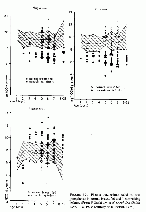
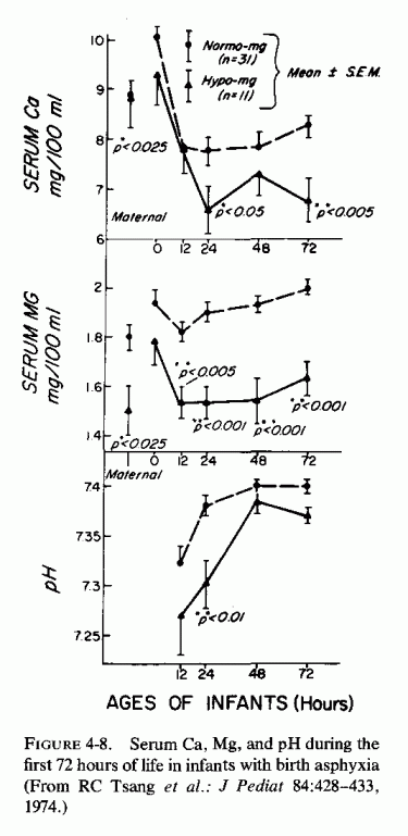
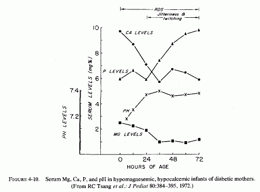
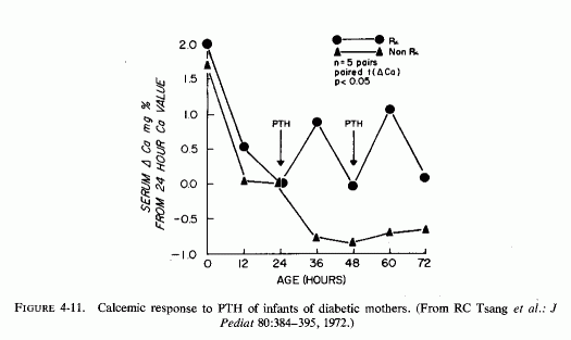
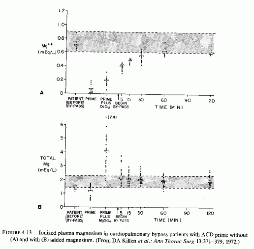
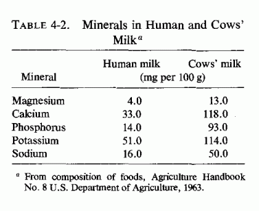
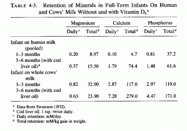
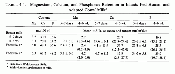
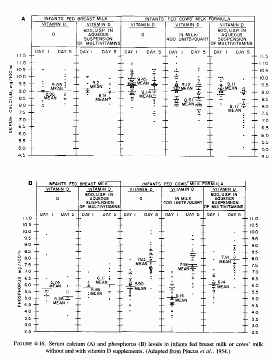
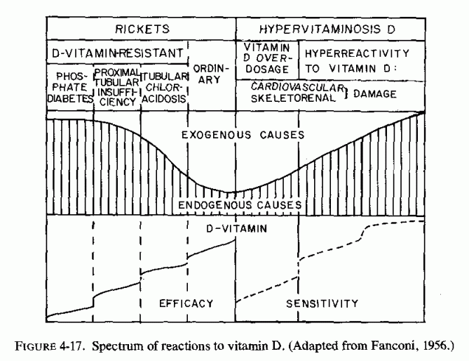
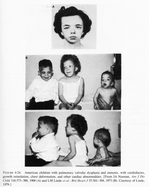
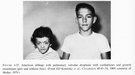
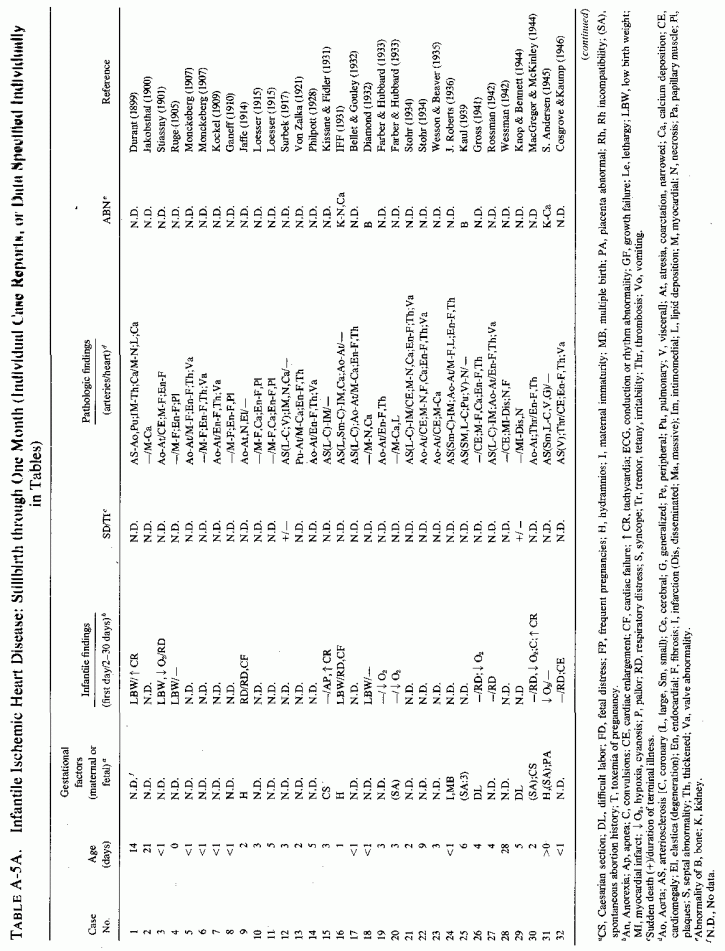
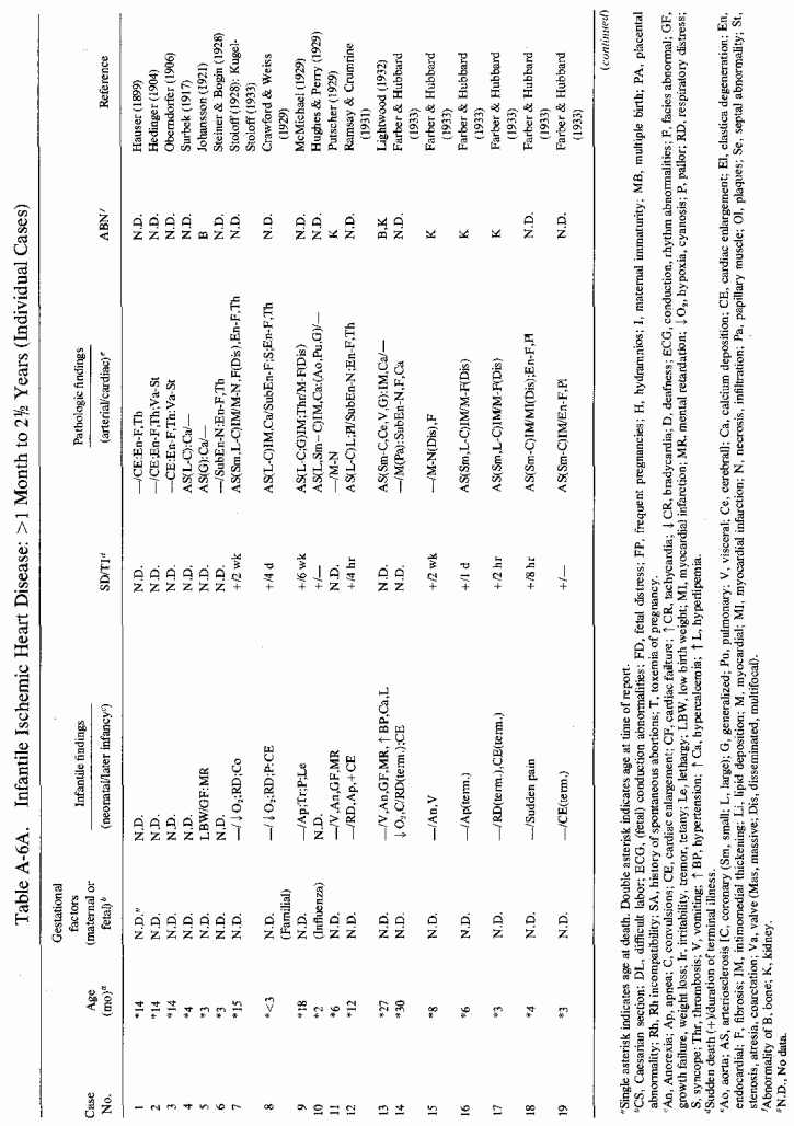